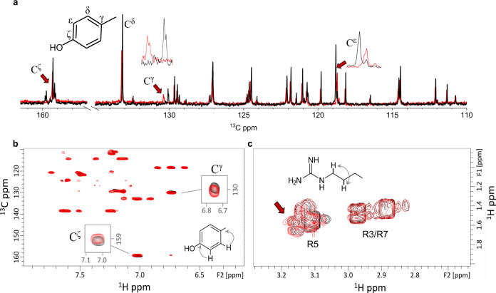
Fig. 6. NMR determines the molecular mechanism of droplet formation.
a The one-dimensional 13C spectrum for the WGR-1 peptide at 20 (black, LLPS) and 5 (red, non-LLPS) mM in 50 mM tris buffer pH 10 and 300 K. Arrows indicate chemical shift differences at the Y14 aromatic ring. b Aromatic region of the 2D-1H,13C-HMBC spectrum showing long-range proton-carbon correlations allowing the detection of quaternary carbons. Y14 chemical shift changes are shown as before. c Region of the 2D-1H, 1H-COSY spectrum showing the correlation between arginine Hγ–Hδ protons.

