
Figure 2.
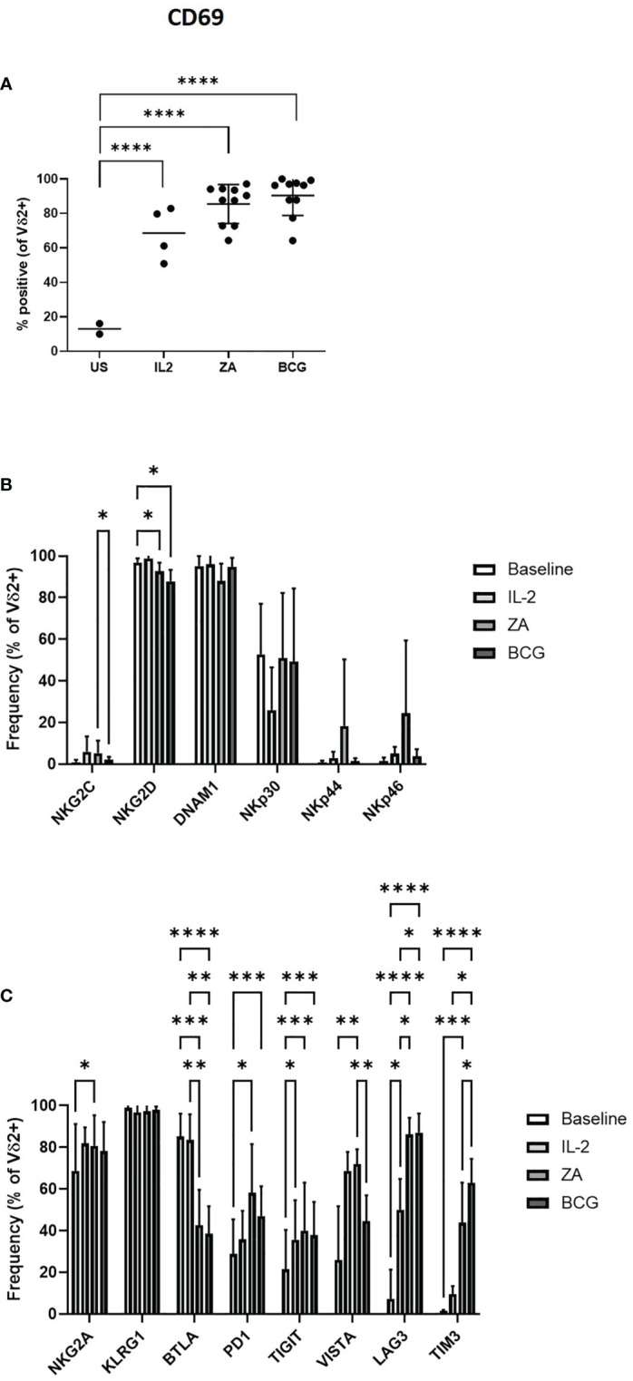
Activation of Vδ2 cells was assessed by flow cytometry of CD69 expression following 24 hours stimulation with IL-2, ZA or BCG both with IL-2 (A). Expression of NK associated activatory markers (B) and inhibitory checkpoint receptors (C) was determined on Vγ9Vδ2 T-cells in PBMC stimulated for 24 hours with IL-2 alone, ZA or BCG, both with IL-2, using flow cytometry. N=10. *p<0.05, **p<0.005, ***p<0.0005, ****p<0.0001, non-parametric mixed effects analysis with Tukey’s post hoc for multiple pairwise comparisons.