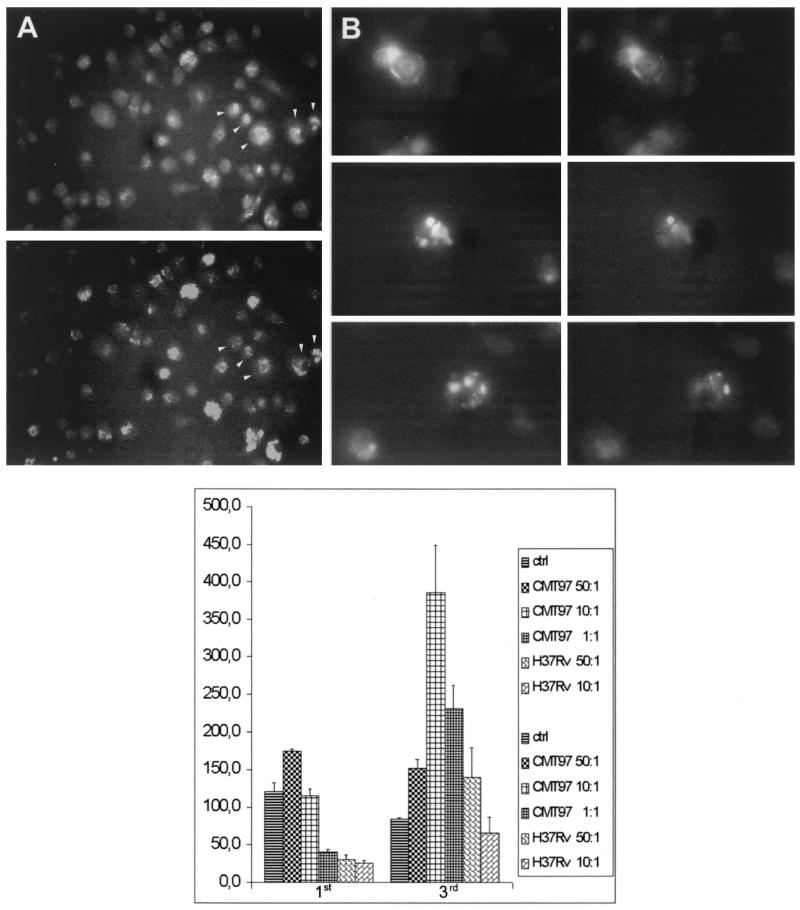
FIG. 2.
(A) Immunofluorescence of H37Rv-infected macrophages. The top panel shows anti-CD14 staining in nonpermeabilized macrophages; the bottom panel shows anti-IFN-γ staining in permeabilized macrophages. The arrowheads indicate macrophages that are positive for both antibodies. (B) Immunofluorescence of H37Rv- and CMT97-infected macrophages. Top and bottom panels, macrophages infected with the CMT97 strain. Middle panel, macrophages infected with the H37Rv strain. The left column shows anti-CD14 staining, and the right column shows anti-IFN-γ staining. (C) ELISA titration of IFN-γ released in culture supernatants of human macrophages infected with H37Rv and CMT97. The macrophages were prepared as described in the text and infected for 3 h at different MOIs (50:1,10:1, and 1:1); after 1 and 3 days of infection, the culture supernatants were collected, and IFN-γ protein was measured. Compared with the laboratory strain H37Rv, clinical strain CMT97 infection induces higher and more prolonged IFN-γ production.

