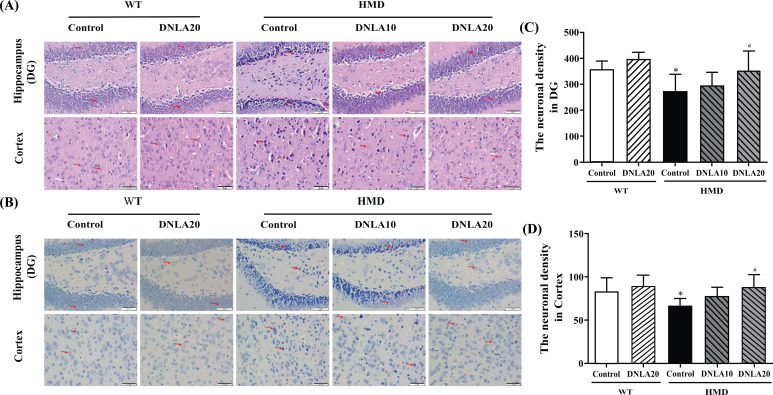
Fig. (2).
Effects of DNLA on morphological alterations in the hippocampus and cortex. Sections of the hippocampal DG region and cortex were obtained and stained with HE and Nissl (magnification, 400×). (A) Representative HE staining cyto-architecture. (B) Representative Nissl staining cytoarchitecture. (C) Statistics of viable neurons in the hippocampal DG region. (D) Statistics of viable neurons in the cortex region. Note: data are mean ± SEM, n = 6, *p < 0.05 vs Control group; #p < 0.05 vs HMD group.

