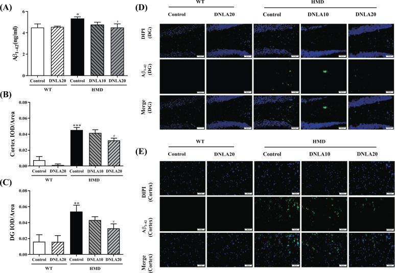
Fig. (6).
Effect of DNLA on Aβ1-42 levels in hippocampal tissue, cortex tissue and serum. Sections of the hippocampus and cortex were obtained and stained with Aβ1-42 (magnification, 400×). (A) The Aβ1-42 level in serum. (B) IOD/Area of Aβ1-42 in the hippocampal DG region. (C) IOD/Area of Aβ1-42 in the cortex region. (D) Representative photomicrographs in the hippocampal DG region. (E) Representative photomicrographs in the cortex region. Note: data are mean ±SEM, n = 6 or 8, *p < 0.05, **p < 0.01, ***p < 0.001 vs. Control group; #p < 0.05 vs. HMD group.

