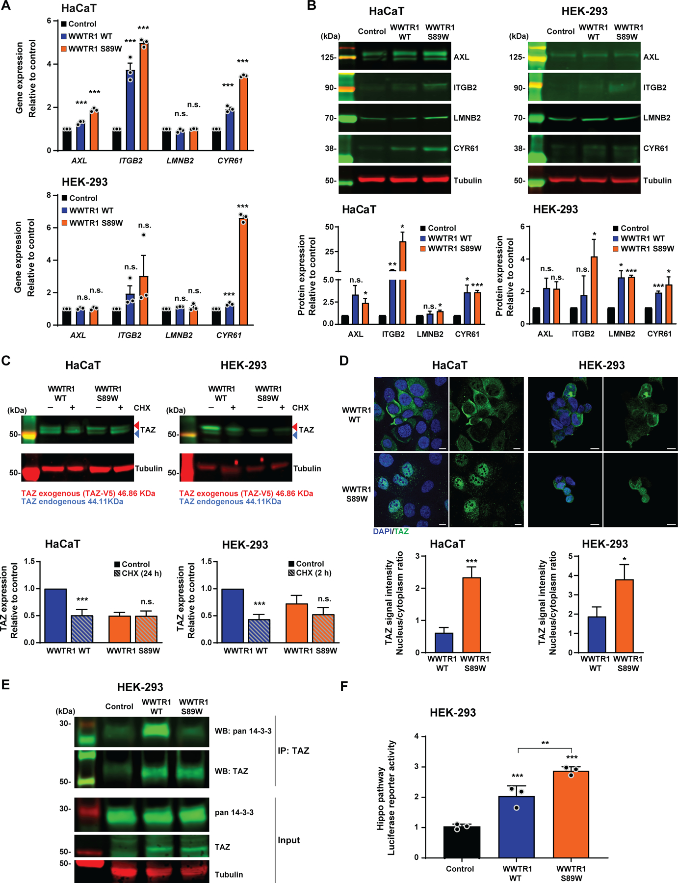
Figure 3. WWTR1 S89W expression results in the overexpression of Hippo pathway targets in vitro and in deregulation of the Hippo pathway.

(A) Quantitative assessment of Hippo pathway targets AXL, ITGB2, LMNB2 and CYR61 transcripts in immortalized human keratinocyte HaCaT and HEK-293 cells stably expressing empty vector (Control), WWTR1 wild-type (WWTR1 WT) and WWTR1 S89W. Expression levels were normalized to GAPDH expression, and comparisons of mRNA expression levels were performed relative to Control. (B) Representative western blot analysis of Hippo pathway targets AXL, ITGB2, LMNB2 and CYR61 protein levels in HaCaT and HEK-293 cells stably expressing Control, WWTR1 WT and WWTR1 S89W. Tubulin was used as protein loading control. Quantification (bottom) of protein levels as compared to Control. (C) Representative western blot analysis of TAZ protein levels in immortalized human keratinocyte HaCaT and HEK-293 cells stably expressing WWTR1 WT and WWTR1 S89W treated with 50 μM Cycloheximide (CHX). Tubulin was used as protein loading control. Quantification (bottom) of protein levels as compared to untreated WWTR1 WT cells. (D) Representative confocal micrographs of immunofluorescence analysis of TAZ (green) and 4–6-diamidino-2-phenylindole (DAPI, blue) in HaCaT and HEK-293 cells stably expressing Control, WWTR1 WT and WWTR1 S89W (scale bars, 50 μm). Quantification (bottom) of TAZ intensity/ cell in a nucleus/cytoplasm ratio. (E) Immunoprecipitation assay with TAZ antibody of HEK-293 cells stably expressing Control, WWTR1 WT or WWTR1 S89W. Western blot was performed using anti-TAZ and anti-pan 14–3-3 antibodies, and tubulin as loading control. (F) Hippo pathway luciferase reporter assay of HEK-293 cells stably expressing Control, WWTR1 WT and WWTR1 S89W. SV40-Renilla was used to normalize transfection efficiency. All experiments were performed in technical triplicate in at least 3 independent replicates and statistical analysis was performed comparing each condition to Control (A,B,F), or to WWTR1 WT (C,D). Mean ± SD; n.s., not significant; *P<0.05, **P<0.01, ***P<0.001; two-tailed unpaired t-test.
