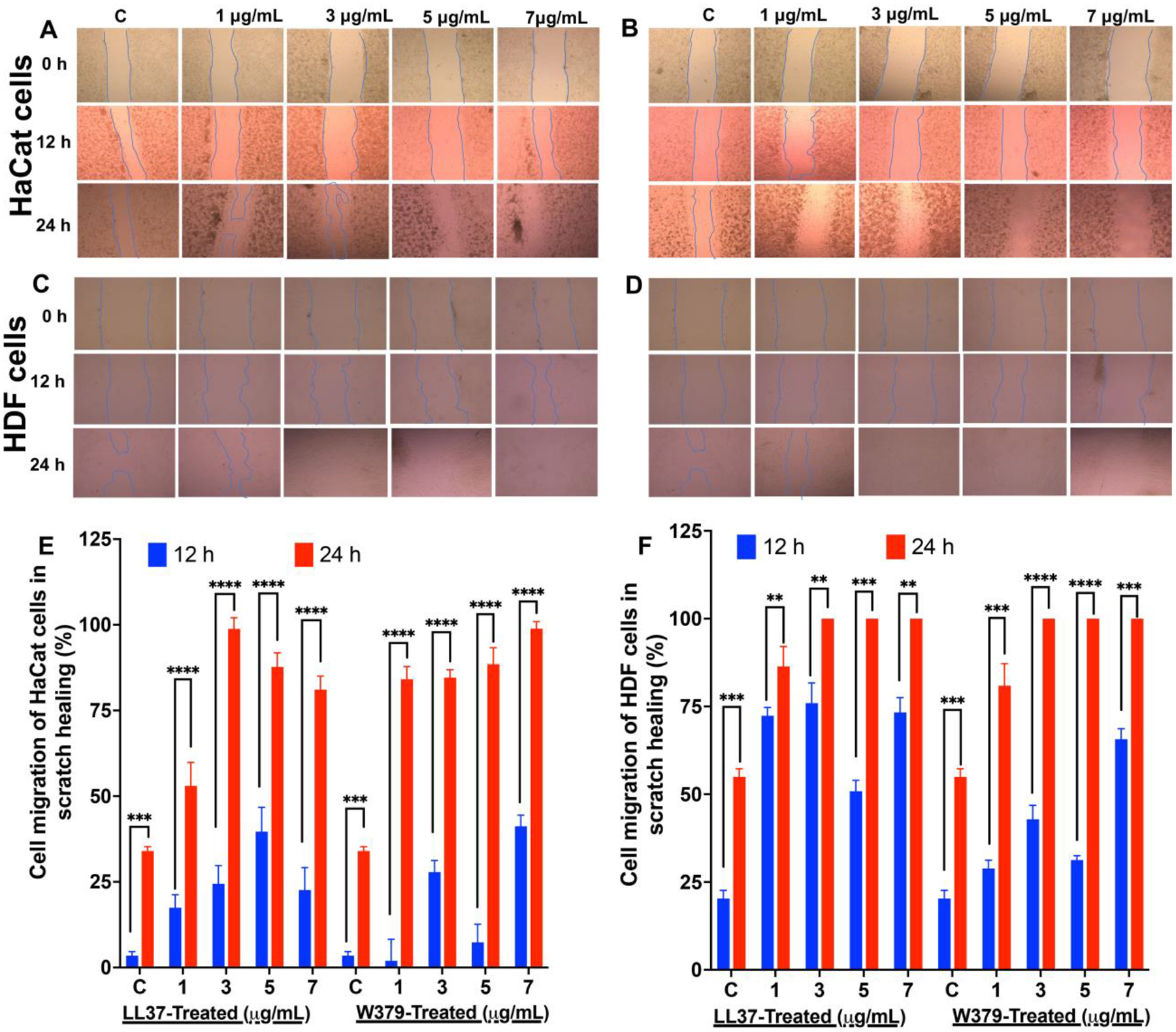
Figure 3. Scratch assay and cell migration analysis.

(A) Representative bright field images show the migration of HaCat cells in the scratch area after treated with different concentrations of W379 peptide. (B) Representative bright field images show the migration of HaCat cells in the scratch area after treated with different concentrations of LL-37 peptide. (C) Representative bright field images show the migration of HDF in the scratch area after treated with different concentrations of W379 peptide. (D) Representative bright field images show the migration of HDF in the scratch area after treated with different concentrations of LL37 peptide. (E) Wound closure expressed in scratch assay of HaCat cells treated with LL37 and W379 peptide at different concentrations ranging from 1 μg/mL to 7 μg/mL. (F) Wound closure expressed in scratch assay of HDF treated with LL37 and W379 peptide at different concentrations ranging from 1 μg/mL to 7 μg/mL. **p<0.01, ***p<0.001, ****p<0.0001.
