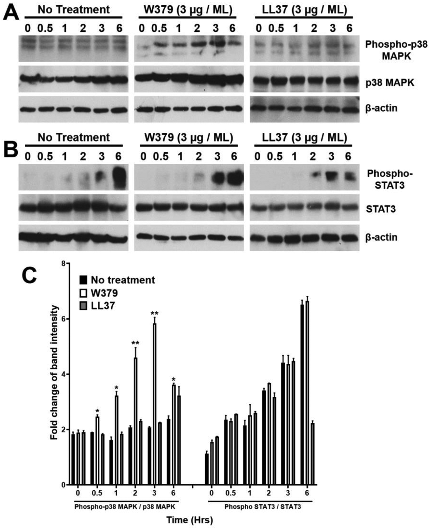
Figure 6. W379 peptide may promote keratinocyte migration via the p38 MAPK signaling pathway.

Total protein from HaCat cells treated with either W379 or LL-37 peptide (3 μg/mL) was analyzed for the phosphorylation status of p38 MAPK and STAT3 at varying time points. Non treated cells collected at respective time points served as control (No treatment). (A) Western blot depicting phospho-p38 MAPK and total p38 MAPK expression in control, W379 and LL-37 peptide treated cells. (B) Protein analysis of STAT3 phosphorylation status in control, W379 and LL37 peptide treated cells. (C) Quantitation of the densitometric analysis of all replicates corresponding to (A) and (B) conducted using Image J. Band intensity of phosphorylated protein to total protein was calculated and the fold change represented as a graph with statistical significance depicted as *p<0.05, **p<0.01.
