
Figure 1. Sample preparation and CyTOF identification of major leukocyte subsets including CD15+ /CD66b+ neutrophils.
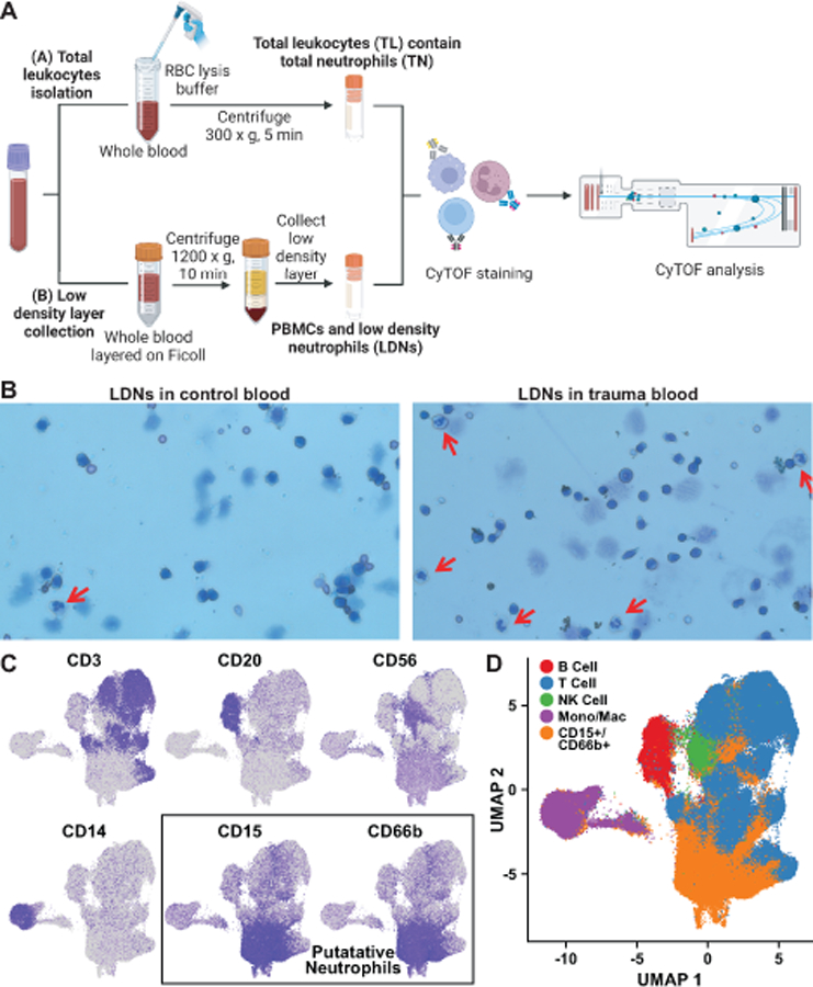
A. Separation procedure for purification of total leukocytes (TL) including total neutrophils (TN), and the PBMC layer including low-density neutrophils (LDN). B. Cells in the PBMC layer were visualized by Wright’s stain. Mature neutrophils are indicated by the red arrows. C, D. Markers including CD3, CD20, CD14, CD15, and CD66b (C) were used to identify clusters of cells as B cells, T cells, monocytes/macrophages, or neutrophils (D). Blue color intensity in individual marker plots indicates level of surface marker detection by CyTOF, log-normalized as described in methods, with the bluest color corresponding to the highest expression of that marker.