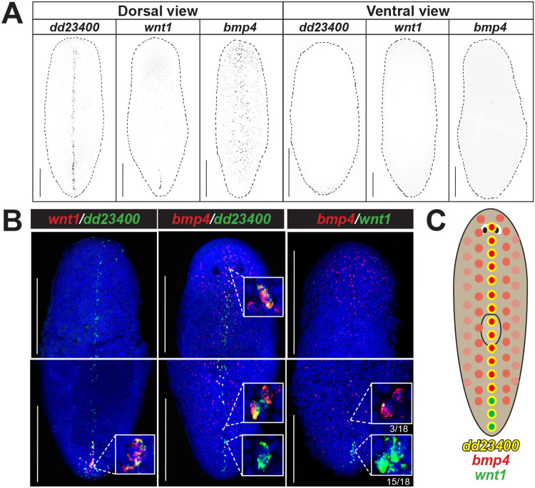
Fig. 1. bmp4 and wnt1 co-express with dd23400+ dorsal midline cells in a regionally distinct manner.
(A) Fluorescent in situ hybridization (FISH) detecting dd23400 on the dorsal midline, wnt1 on the dorsal posterior midline, and bmp4 in a dorsal midline-centered gradient with reduced expression in the far posterior. Scale bars represent 150 μm with dorsal or ventral views indicated. (B) Double FISH detecting co-expression of wnt1, dd23400, and bmp4. 88.9% of wnt1+ cells co-expressed dd23400 and 55.4% of dd23400+ cells co-expressed bmp4. Only 12.1% of wnt1+ cells co-expressed bmp4. Therefore, along dd23400+ dorsal midline muscle cells, wnt1 and bmp4 expression defines largely nonoverlapping AP domains within posterior. Within the wnt1+ domain of the representative image, wnt1+ cells in the anterior co-expressed bmp4 (3/18) whereas cells in the posterior of the domain lacked bmp4 co-expression (15/18). Scale bars represent 150 μm. (C) Schematic illustrating separation of bmp4 and wnt1 domains on the dorsal midline.

