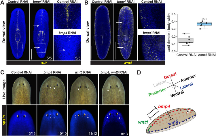
Fig. 4. bmp4 regulates ML patterning upstream of lateral wnt5.
(A-B) FISH detecting slit or wnt5 following 28 days of homeostatic control or bmp4 RNAi. Right panels show enlargements of boxed regions. Scale bars represent 300 μm. (A) bmp4 RNAi caused reduction of slit in the anterior and elimination in the posterior (arrows). (B) Inhibition of bmp4 caused medial expansion of wnt5 (arrows). Graph shows wnt5 expression domain width (dorsal side) normalized to body width. ****p<0.0001 by unpaired 2-tailed t-test; N ≥ 6 animals. Box plots show median values (middle bars) and first to third interquartile ranges (boxes); whiskers indicate 1.5× the interquartile ranges and dots are data points from individual animals. (C) Eyes assessed by live imaging (top) or opsin FISH (bottom) after 21 days of homeostatic RNAi to inhibit bmp and/or wnt5 and scored for lateral or medial ectopic eyes (arrows). For single-gene RNAi, dsRNA for the targeted gene was mixed with an equal amount of control dsRNA so that the total amounts of each targeted dsRNA delivered were equal between single- and double-RNAi conditions. 100% of bmp(RNAi) animals had ectopic medial eyes and 92% wnt5(RNAi) animals had ectopic lateral eyes. By contrast, 100% of bmp4;wnt5(RNAi) animals had at least two lateral ectopic eyes, and of these 38% also formed a single medial ectopic eye while 62% only formed ectopic lateral eyes (shown). Therefore, inhibition of wnt5 masks the bmp4(RNAi) ML phenotype. (D) Model of bmp4 establishing DV polarity, influencing AP regulation through wnt1, and controlling ML patterning through wnt5.

