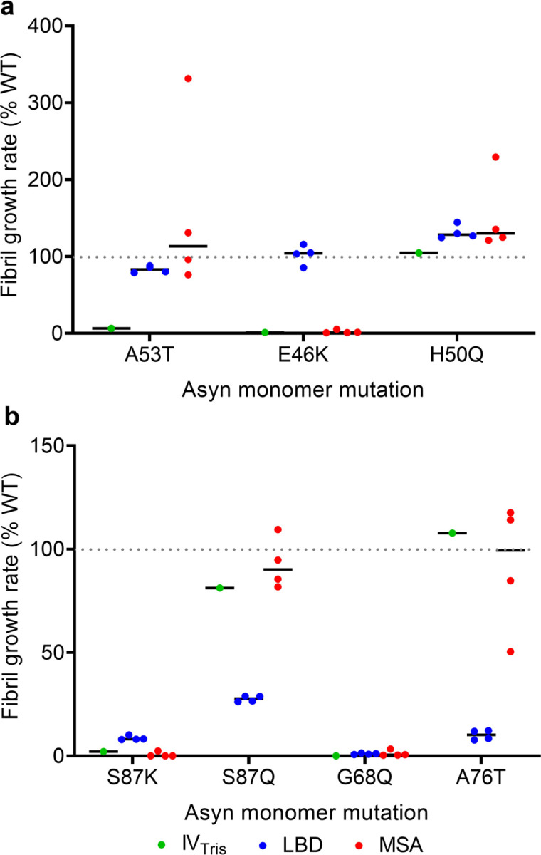
Fig. 6: Fibril growth rates for in vitro assembled fibrils and amplified fibrils in the presence of mutant Asyn monomer.

a, WT-normalized growth rates in the presence of Asyn monomer with familial PD mutations A53T, E46K and H50Q. b, WT-normalized growth rates in the presence of Asyn monomer with S87K, S87Q, G68Q and A76T mutations. The results show that the A53T mutation distinguishes in-vitro (Tris buffer) fibrils from the LBD and MSA amplified fibril forms and that the E46K-mutation distinguishes MSA amplified fibrils from LBD amplified fibrils. The S87Q and A76T mutations distinguish LBD amplified fibrils from in-vitro and MSA amplified fibril forms.
