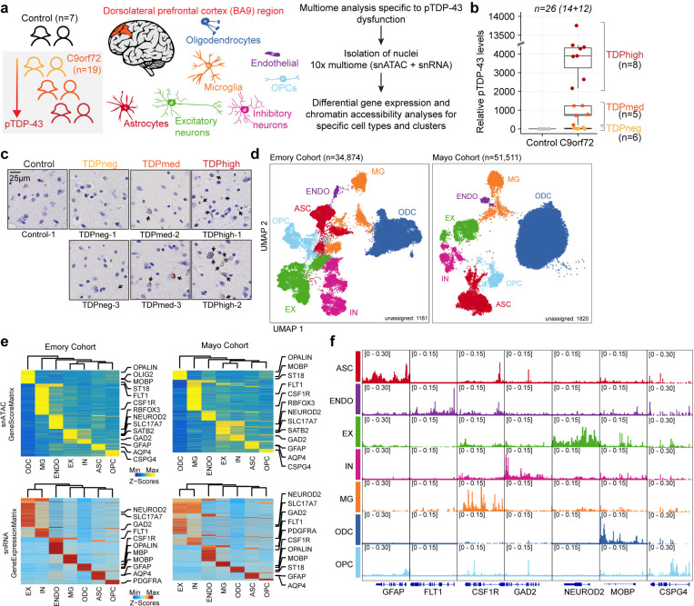
Fig. 1. Multiomic single-nucleus analyses identify diverse cortical cell types in the dorsolateral prefrontal cortex of controls and C9orf72 ALS/FTD patients with different levels of pTDP-43.
(a) Schematic representation of single-nucleus multiome profiling (snATAC-seq and snRNA-seq in the same nuclei) of dorsolateral prefrontal cortex samples from 7 control and 19 c9orf72 ALS/FTD donors analyzed in this study. (b) pTDP-43 levels in control and C9orf72 ALS/FTD patient cortical tissues. (c) Evaluation of the presence of pTDP-43 aggregates in C9orf72 ALS/FTD patient cortical tissues and controls. (d) snATAC-seq and snRNA-seq integrated UMAP visualization of major cortical cell types in samples from the Emory (left) and Mayo (right) cohorts, where each dot corresponds to each of the nuclei profiled simultaneously for transcriptome and chromatin accessibility using the 10x multiome platform. (e) Row-normalized single-nucleus gene expression (top) or gene score (bottom) heatmaps of cell-type marker genes for Emory (left) and Mayo (right) cohorts. (f) Pseudo-bulk chromatin accessibility profiles for each cell type at cell-type marker genes in the Emory cohort.

