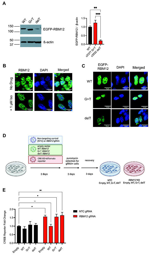
Figure 4. Expression of disease-associated variants in RBM12 knockdown cells does not rescue the hyperactive GPCR-dependent transcriptional signaling.

(A) Western blot analysis of EGFP-tagged wild-type, c.2377G>T and c.2532delT RBM12 (n = 3) probed with antibody recognizing EGFP. All data are normalized relative to wild-type. (B) Fixed cell fluorescence microscopy analysis of endogenous RBM12 in untreated and stimulated (1 μM Iso for 20 minutes) HEK293 cells. (C) Localization of EGFP-tagged wild-type, c.2377G>T or c.2532delT RBM12 by fluorescence microscopy. (D) Schematic of the flow cytometry-based rescue experiment. (E) Flow cytometry measurement of the fluorescent CREB transcriptional reporter (CRE-DD-tdTomato) in response to 1 μM Iso and 1 μM Shield for 6 hours (n = 8-11). Data are normalized relative to the “NTC + empty vector” sample values. All data are mean ± SEM. Statistical significance was determined using one-way ANOVA (A) or two-way ANOVA with Dunnett’s correction (E). See also Figure S4.
