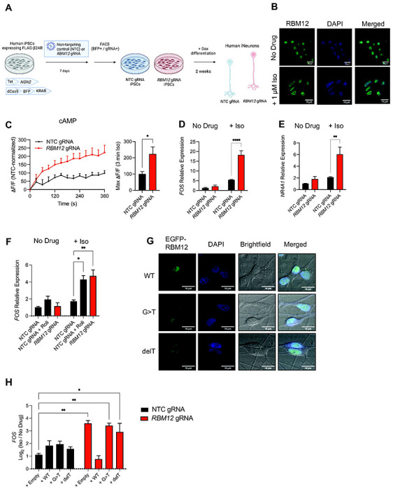
Figure 5. Signaling hyperactivity upon loss of RBM12 in human iPSC-derived neurons.

(A) Schematic of the CRISPRi-mediated RBM12 depletion in human iPSC-derived neurons. (B) Fixed cell fluorescence microscopy imaging of native RBM12 in untreated and stimulated (1 μM Iso for 30 minutes) iNeurons. (C) Accumulation of the fluorescent cADDis sensor in NTC- and RBM12 gRNA-expressing neurons in response to 1 μM Iso (n = 6). (D) Expression of FOS and (E) NR4A1 mRNAs in response to treatment with 1 μM Iso for 1 hour by RT-qPCR (n = 6). (F) Expression of FOS mRNA in response to 1 μM Iso in the presence of either vehicle (DMSO) or 10 μM Rolipram in neurons treated for 1 hour (n = 3). (G) Expression of EGFP-tagged wild-type, c.2377G>T or c.2532delT RBM12 by fluorescence microscopy. (H) Expression of FOS mRNA in response to stimulation with 1 μM Iso for 1 hour in neurons transduced with empty plasmid or plasmid encoding WT, G>T, or delT EGFP-RBM12 (n = 2-3). All data are mean ± SEM. Statistical significance was determined using unpaired t-test (C) or two-way ANOVA with Tukey’s correction (D-F, H). See also Figure S5.
