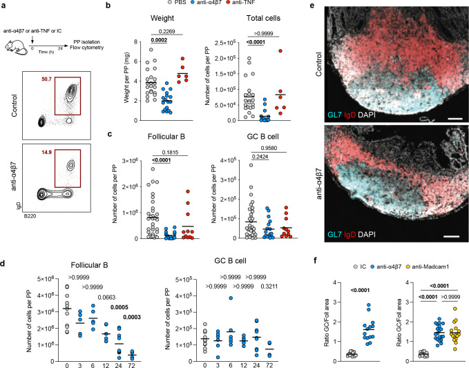
Figure 3. Anti-α4β7 antibody administration results in the attrition of Peyer’s patches in mice.
a, Mice received one intraperitoneal injection of anti-α4β7 antibody, anti-TNF antibody or PBS antibody, and were sacrificed 24 hours later. Cells from PPs were analyzed by flow cytometry to quantify follicular B cells (CD45+B220+IgD+) and germinal center B cells (CD45+B220+IgD−GL7+FASL+). b, Weight and total cell counts from individual PPs taken 24h after injection. c, Frequency of follicular B cells and germinal center B cells from individual PPs. d, Frequency of follicular B cells and germinal center B cells from PPs of untreated mice (grey) and at 3, 6, 12, 24 and 72 hours after injection. e, Representative immunofluorescence images from PPs stained for IgD (red), GL7 (cyan) and DAPI (grey), from mice treated with anti-α4β7 antibody or Isotype control. Scale bar indicates 100μm. f, Ratio of germinal center area (GL7+) and follicular area (IgD+) from mice treated with anti-α4β7 (blue), anti-MAdCAM-1 (yellow) or isotype control (gray) antibody. Data shown as individual values and mean. Unpaired non-parametric analysis was done using Kruskal-Wallis test and Dunn’s multiple comparisons test. The p values are indicated.

