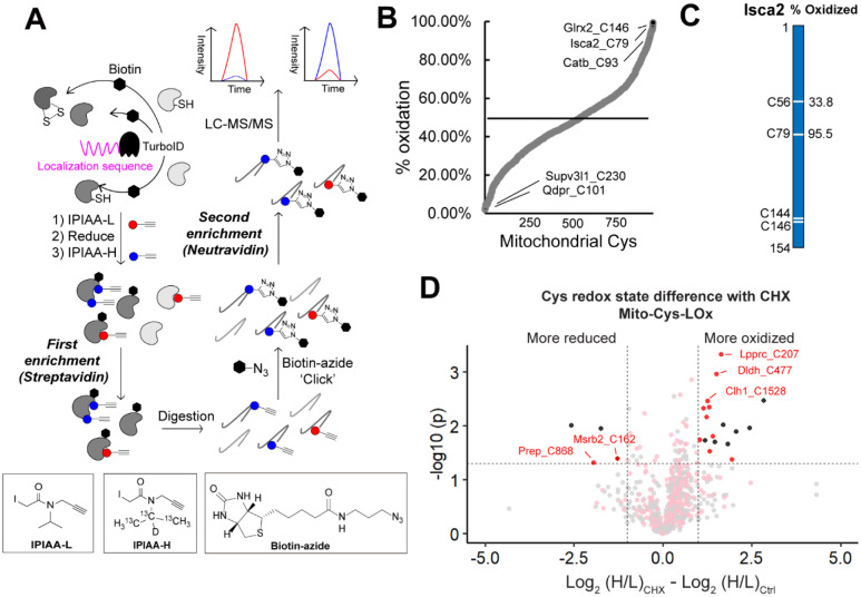
Figure 4. Establishing the Local Cysteine Oxidation (Cys-LOx) method to analyze basal mitochondrial cysteine oxidation states.
A) Scheme of Cys-LOx method. B) Percent oxidation state of mitochondrial cysteines identified with mito-Cys-LOx. C) Percent oxidation state of cysteines quantified in an exemplary mitochondrial protein. D) Difference in redox states of cysteines quantified with Mito-Cys-LOx with or without dialyzed FBS and CHX treatment. Red dots indicate cysteines localized in mitochondria. Black dots indicate cysteins localized in organelles other than mitochondria. Dialyzed FBS (Dia-FBS) treatment was 36 h and CHX treatment was 100 ug/mL for 6 h at 37 °C. Experiments were performed in triplicate in iBMDM cells. All MS data can be found in Table S4.

