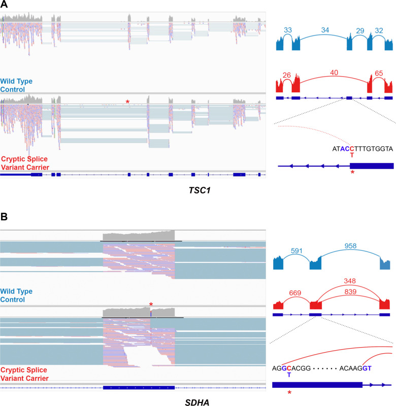
Figure 5. Example cryptic splice variants in established RCC risk genes.
Left: IGV screenshot of the tumor mRNA sequencing data of the wildtype control (above) and the carrier of cryptic splice variant (bottom). Right: Sashimi plot showing the pattern of splicing with the numbered split junction reads.
A) Disruption of splice donor motif led to complete exon skipping in TSC1
B) The cryptic splice variant in SDHA introduced a new splice donor motif GT inside an exon

