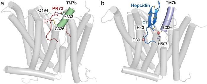
Fig 8. Structural comparison of PR73 and hepcidin-bound HsFpn.
Side views of HsFpn-PR73 (a) and HsFpn-Hepcidin (6WIK) (b) highlight the disulfide bridge and metal ion coordination. In (a), TM7b is colored pale green and PR73 in brick red. In (b), TM7b is colored in light blue, hepcidin shown as marine cartoon with cysteine residues, and C-terminal carboxylate shown as sticks.

