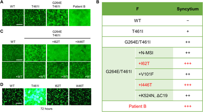
Fig. 4. The F fusogenicity of the patient B strain.
(A) The WT H protein, MeV F [WT F, F(T461I), F(G264E/T461I), and Patient-B-F], CADM1, and EGFP were expressed in 293FT cells. The cells were observed 24 hours after transfection under a fluorescence microscope. Scale bar, 500 μm. (B) The F mutations that the patient B strain has and the fusogenicities of F proteins having certain combinations of the mutations are shown. N-MSI indicates three amino acids (methionine, serine, and isoleucine) added at the N terminus. ΔC19 indicates a 19–amino acid deletion at the C terminus. (C) The WT H protein, MeV F [WT F, F(G264E/T461I), F(I62T/G264E/T461I), or F(G264E/T461I/I446T)], CADM1, and EGFP were expressed without or with the WT F protein in 293FT cells. The cells were observed 24 hours after transfection under a fluorescence microscope. Scale bar, 500 μm. (D) The WT H protein, MeV F [WT F, F(T461I), F(I62T), or F(I446T)], CADM1, and EGFP were expressed in 293FT cells. The cells were observed 72 hours after transfection under a fluorescence microscope. Scale bar, 500 μm.

