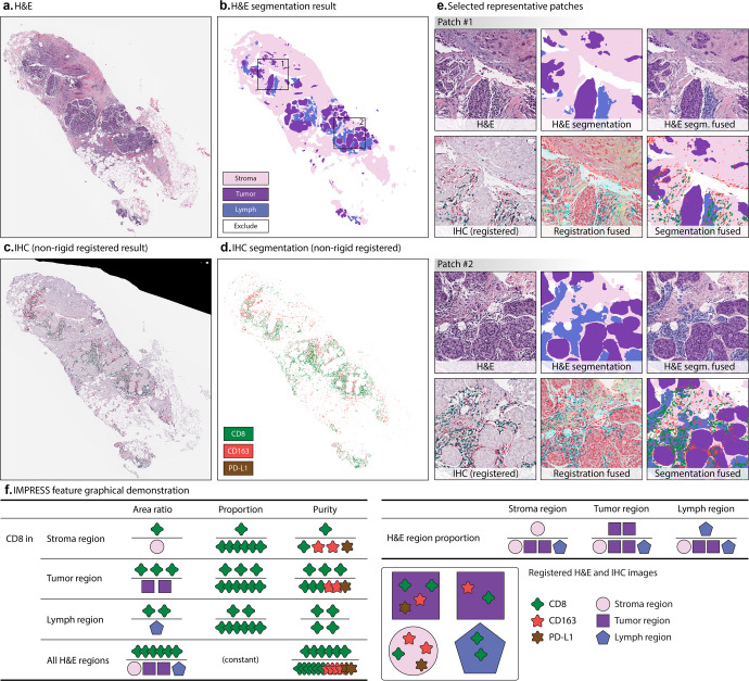
Fig. 2. Tissue segmentation and image-level features extraction from registered H&E and IHC segmentation.
a An example H&E tissue; b H&E tissue segmentation result; c IHC tissue (aligned to a) after non-rigid registration; d IHC segmentation results, after non-rigid registration. e Selected representative patches from b including (1) H&E patch, (2) H&E segmentation, (3) H&E segmentation (segm. in short) fused with original patch, (4) IHC patch after registration, (5) IHC patch after registration fused with H&E patch, and (6) H&E, IHC segmentation fused patch; f IMPRESS feature graphical demonstration. In f, each IHC marker produces 11 features (CD8 was shown as an example), H&E region produces 3 features, totally 36 IMPRESS features. Figure best viewed in color.

