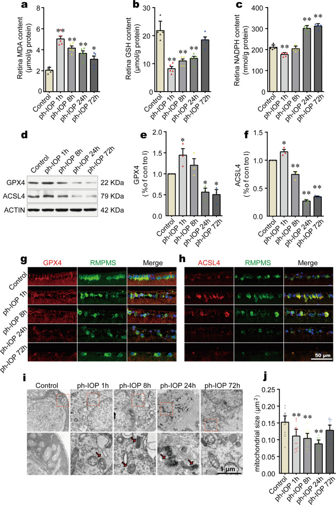
Fig. 3. Pathologically high intraocular pressure (ph-IOP)-induced iron accumulation led to retinal ferroptosis at the early phase.
Comparison of MDA (a), GSH (b), and NADPH (c) contents between control mice and ph-IOP injured mice (n = 5 in each group). d–f Western blotting detection of retinal levels of GPX4 and ACSL4 (normalized to that of actin) in control and ph-IOP injured mice (n = 3 in each group). Representative photomicrographs of immunofluorescence staining for GPX4 (g; red) and ACSL4 (h; red) in retinal ganglion cells (counterstained with RBPMS; green). i Representative photomicrographs of transmission electron microscopy showing the mitochondria with ferroptosis features (red arrows) after ph-IOP injury. j Comparison of mitochondrial size between control mice and ph-IOP injured mice (n = 15 in control, 1 h and 72 h groups; n = 14 in 8 h and 24 h groups). MDA malondialdehyde, GSH, glutathione, NADPH nicotinamide adenine dinucleotide phosphate, GPX4 glutathione peroxidase 4, ACSL4 Acyl-CoA synthetase long-chain family member 4, RBPMS RNA-binding protein with multiple splicing. Data are shown as the mean ± SD; *p < 0.05, **p < 0.01 (compared with the control group using one-way analysis of variance). Bar = 50 μm (g and h) and 1 μm (i).

