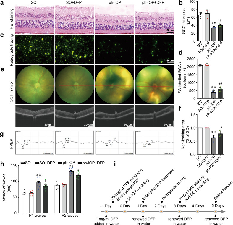
Fig. 6. Inhibiting ferroptosis using deferiprone (DFP) protected retinal ganglion cells (RGCs) from pathologically high intraocular pressure (ph-IOP) injury.
Representative photomicrographs of hematoxylin and eosin (HE)-stained retinal slices (a) and the thickness of the ganglion cell complex (GCC; b) in the sham operation (SO) mice and ph-IOP injured mice, with or without DFP treatment (n = 5 in SO groups and n = 7 in ph-IOP groups). Representative photomicrographs of fluorogold (FG)-labeled RGCs from the peripheral retinas (c) and the number of FG-labeled RGCs (d) (n = 6 in SO groups and n = 8 in ph-IOP groups). Representative photomicrographs of optical coherence tomography (OCT)-detected retinas (e) and non-leaking areas (f) (n = 4 in SO groups and n = 6 in ph-IOP groups). Representative photomicrographs of flash visual-evoked potentials (FVEPs; g) and the latency of P1 and P2 waves (h) (n = 6 in SO groups and n = 8 in ph-IOP groups). i Schematic diagram of the verification of the protective effect of DFP on ph-IOP-induced RGC damage. Data are shown as the mean ± SD; **p < 0.01 (compared with the SO group using one-way analysis of variance); #p < 0.05, ##p < 0.05 (compared with the ph-IOP group using one-way analysis of variance). Bar = 50 μm (a and c), 200 μm (e) and 50 ms (g).

