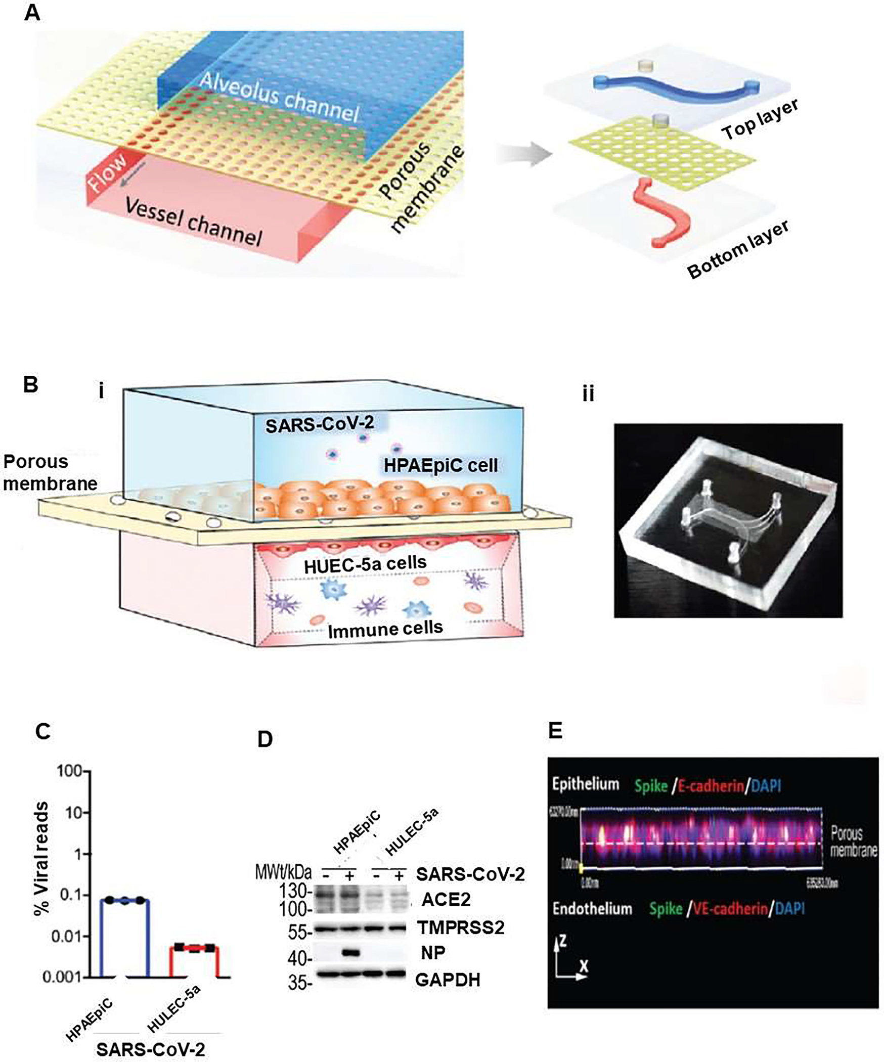
Figure 3.

Human alveolar chip to recapitulate lung injury and immune responses induced by SARS-CoV-2 in vitro. (A) Sschematic diagram represents configuration of human alveolus chip infected by severe acute respiratory syndrome corona virus disease-2 (SARS-CoV-2). The device is divided into regions by a porous PDMS membrane: upper alveolar epithelial channel (blue) and lower pulmonary microvascular endothelial channel (red). (B) Illustration of the chip, which is composed of alveolar epithelial cells (HPAEpiC) and pulmonary microvascular endothelial cells (HULEC-5a) separated by a porous membrane (i).. Human immune cells were infused into the bottom vascular channel. Alveolar chamber was exposed to SARS-CoV-2. (ii) Image of the chip. The response of distinct cell types to the virus were analyzed by: (C) RNA-sequencing (RNA-seq) and (D) western blot Analysis of ACE2, TMPRSS2, and viral nucleoprotein (NP) expression levels in mock- or SARS-CoV-2-infected cells at day 3 after SARS-CoV2 infection. (E) Side view of 3D reconstructed confocal image of human alveolar-capillary-barrier 3 after SARS-CoV-2 infection, which showed that virus was predominantly identified in epithelial layer by viral Spike protein expression. Reproduced from [31], which is an open access article distributed under the terms of the Creative Commons CC BY license.
