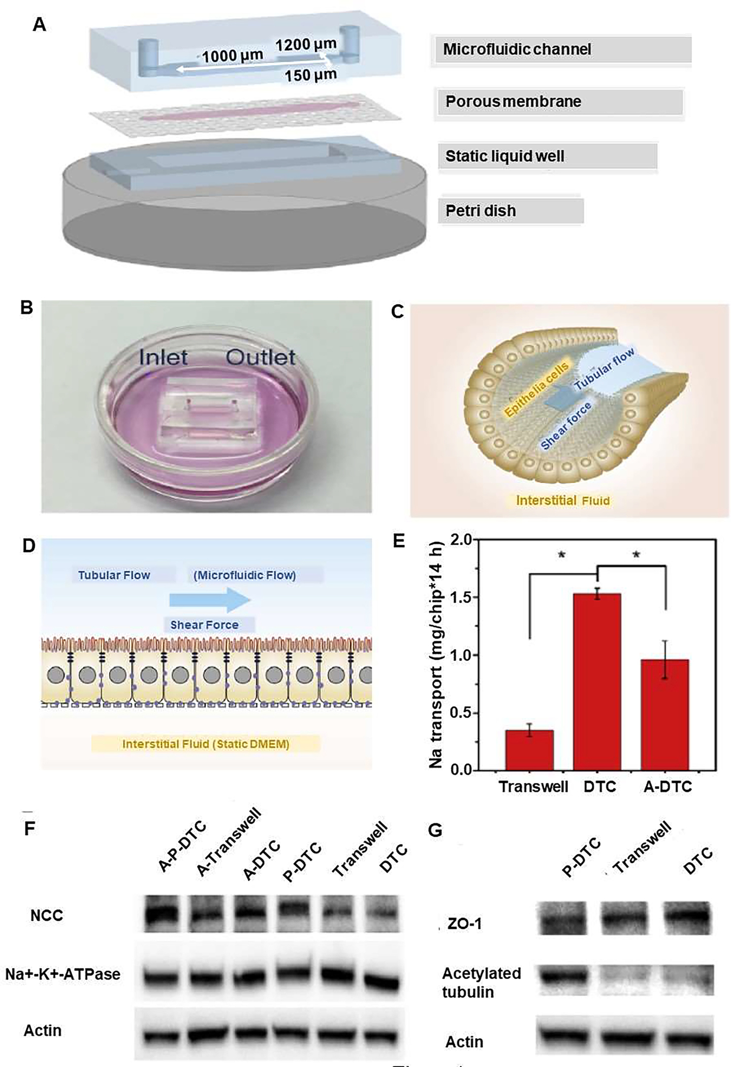
Figure 4.

Distal tubule-on-a-chip (DTC) induced by a Pseudorabies virus (PrV) to investigate the pathogenesis of virus-related renal dysfunctions in electrolyte regulation. Schematic diagram of kidney viral infection-on-a-chip, with inlet and outlet on the top of the microfluidic channel. (A) The three-layered microfluidic chip include 1) microfluidic channel, 2) Madin Darby Canine Kidney (MDCK) cells, epithelial cells from distal renal tubules cultured on a a porous membrane (cell area is shown with pink color), and 3) a static liquid well. The three components were sealed together and placed into a petri dish. (B) A petri dish including media and distal tubule-on-a-chip with inlet and outlet on the top of the microfluidic channel. (C) Photograph of epithelial cells in distal renal tubules and their alignment with the direction of tubular flow, which generated a shear force. (D) Darby Canine Kidney cells were cultured in the chip. The microfluidic flow generated a shear force akin to flowing pro-urine in vivo is created by the tubular flow, and after 24h, infection with PrV (with capsid protein VP26 fused with eGFP started from the luminal side. Before PrV infection, Na reabsorption function was reproduced in DTCs for the tight reabsorption barrier, the microvilli on the apical (luminal sided) membrane, and the polarized-distributed Na transporters. (E) Na reabsorption in DTCs, P-DTCs and A-P-DTCs. (F) Western Blot analyses of NCC and Na+-K+-ATPase in DTCs, Transwell chips, P-DTCs, A-DTCs, A-Transwell chips and A-P-DTCs (G) Western blot analyses of ZO-1 and acetylated tubulin in DTCs, Transwell chips and P-DTCs. Reproduced from [13], with permission from Elsevier.
