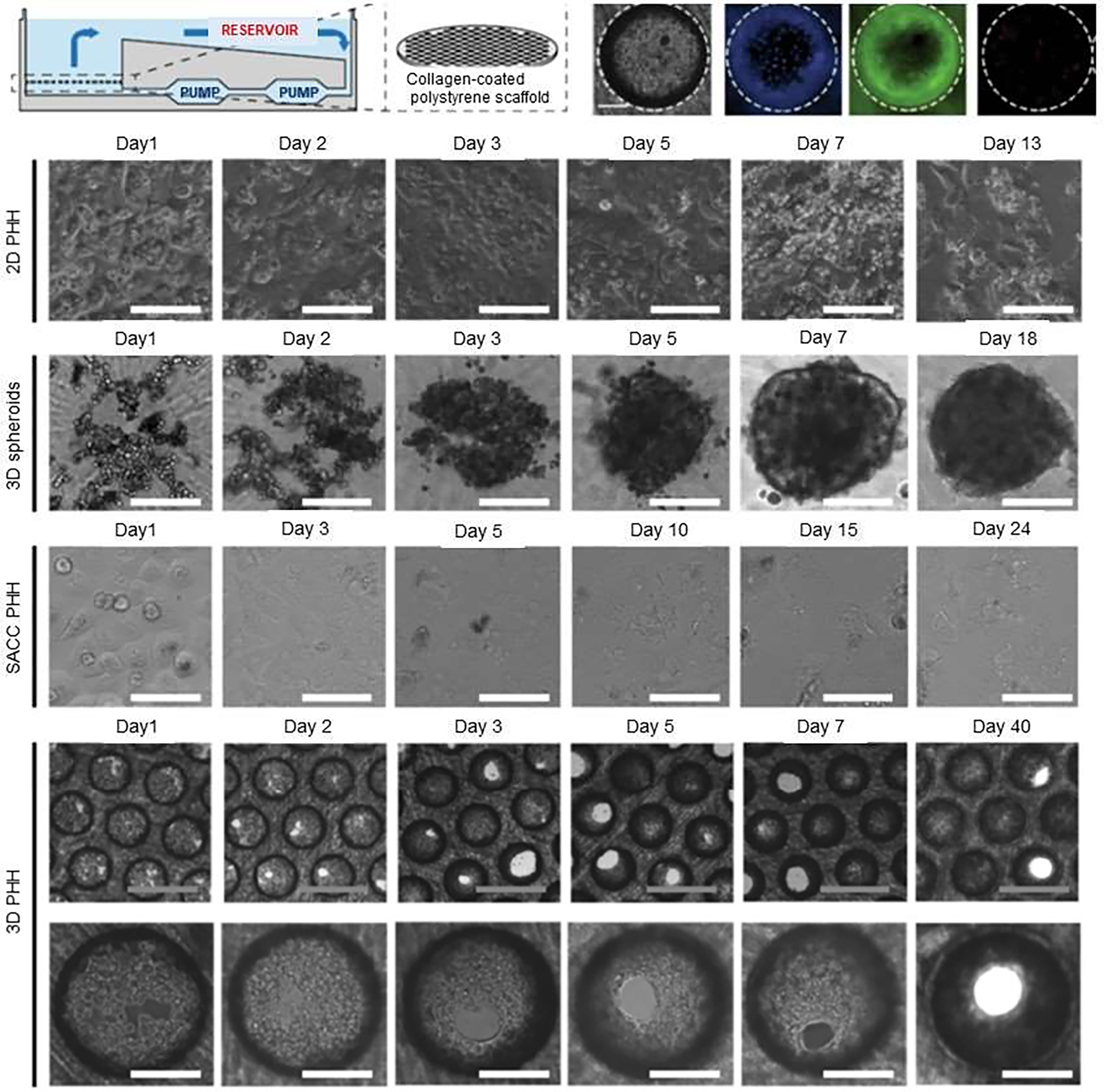
Figure 7.

3D microfluidic liver-on-a-chip having co-cultured primary human hepatocytes (PHHs) with non-parenchymal cells for studying hepatitis B virus (HBV) infection. Formation of physiological hepatic microtissues by three-dimensionally (3D) culturing primary human hepatocytes (PHHs). The schematic represents the perfused bioreactor, initiated by the circulation of media via a pneumatically driven micro-pump, and the application of a collagen-coated scaffold for cell adherence. Cell viability of PHH followed seeding in 3D construct after 13 days post-seeding. Kinetics of hepatic microtissue formation and compared morphologies with 3D spheroid, static two-dimensional (2D) PHH, and self-assembling co-cultures of PHH (SACC) are shown. Immunofluorescence microscopy of albumin (green) and DAPI (blue) in 2D PHH, 3D spheroid, SACC PHH, and 3D PHH cultures after 14 days post-seeding. Reproduced from [57], which is an open access article licensed under a Creative Commons Attribution 4.0 International License.
