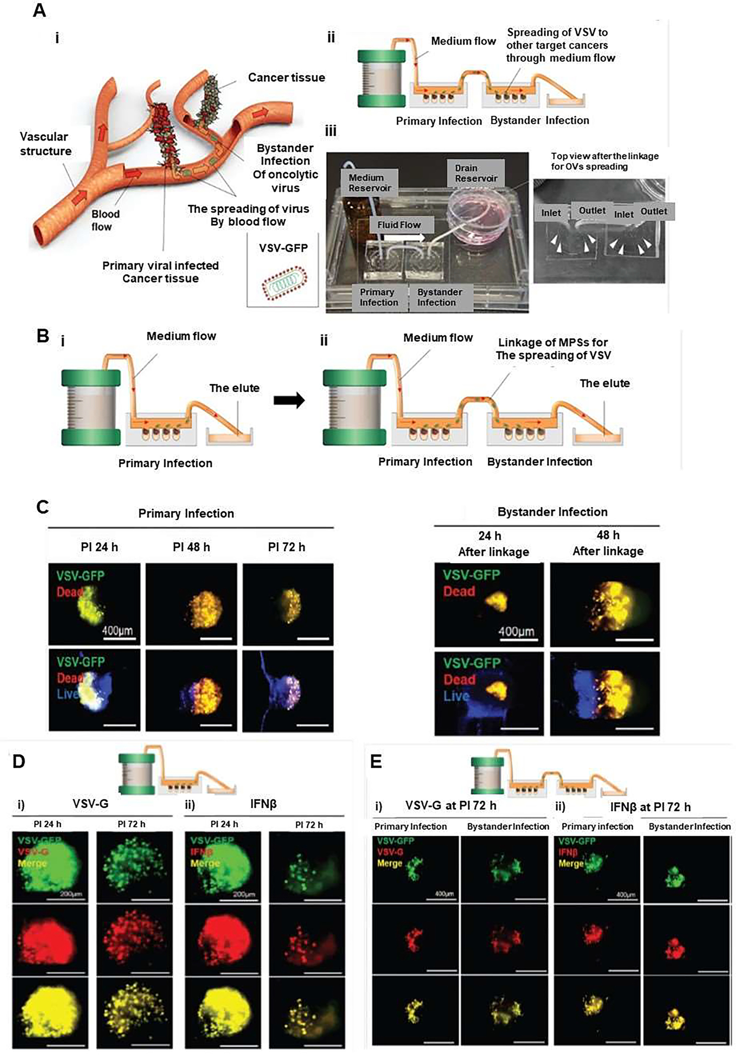
Figure 9.

Three-dimensional (3D) in vitro microphysiological system (MPS) with integrated medium flow to investigate oncolytic viruses (OVs) and bystander infection of OVs. (A) Schematic illustrating how this infection may occur in vivo (i), microphysiological (MPS) model for detecting both oncolytic infection and bystander infection by oncolytic virus spread (ii). (B) The bystander infection of oncolytic viruses by linkage of 3D in vitro MPS by an experimental procedure. (C) The oncolytic effect of the bystander infection of VSV-GFP was assessed by cell death in oncolytic infection and bystander infection of VSV in the MPS with the link system, which confirmed a similar pattern in the linked MPS. (D) The fluorescence expression of VSV-G (i) and IFNβ (ii) proteins in MCTs within MPS of the no-link system at PI 24 and 72 h. (E) Fluorescence expression of VSV-G (i) and IFNβ (ii) proteins in MCTs within MPS of the link system for the bystander effect of VSV-GFP at PI 72 h, which confirmed PI time-dependent fluorescence changes in the primary infected MPS. Reproduced from [65], which is an open access article distributed in accordance with the Creative Commons Attribution (CC BY) license.
