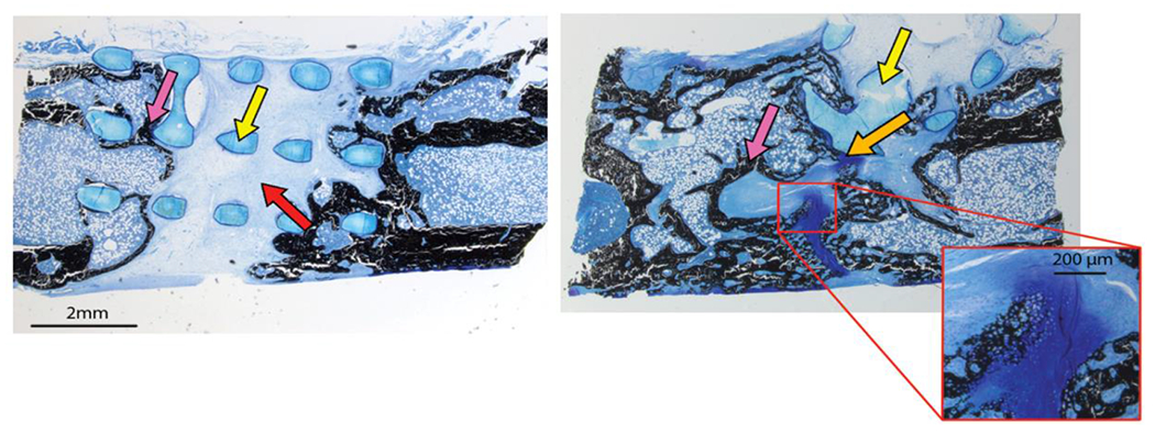
Figure 85.).

VonKossa MacNeil stained longitudinal sections of the femur defect area at 12 weeks. A sample bridged partially by fibrous tissue (left). A sample bridged by combination of bone and cartilage (right). The poly(propylene fumarate) scaffold stains light blue (indicated by yellow arrows), mineralized tissue stains black (indicated by pink arrow), cartilaginous material stains blue/purple (indicated by orange arrows) and fibrous tissue stains a light blue (red arrow). At the 12 week timepoint there was no evidence of the thiol-ene hydrogel macroscopically or microscopically. Endochondral ossification of cartilage is highlighted in the expanded view of the sample on the right. Reproduced, with permission, from reference 426. Copyright 2019 John Wiley and Sons.
