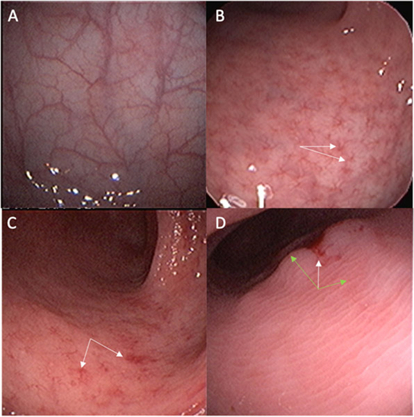
Figure 2. Representative Endoscopic Findings Illustrating the Mayo Score of Endoscopic Severity of Disease.
Representative photos of sigmoidoscopic findings observed (same magnification):
A) Normal mucosa with intact vascular pattern
B) Mild disease with an altered, star-like vascular pattern (white arrows)
C) Mild disease with patchy areas of erythema and mild friability (white arrows)
D) Moderate disease with mucosal edema obscuring all vascular markings (green arrows), with some erythema and friability (white arrow)

