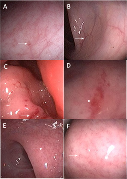
Figure 3. Select Pre-Post Treatment Images of Gastrointestinal Mucosal Changes after 6 Months of Follow-up.
Improvement of mucosal changes: Pre-treatment image (A) showing star-like vascular pattern with mild erythema and post-treatment image (B) showing resolution of abnormalities and new normal mucosa in the same individual.
Worsening of mucosal changes: Pre-treatment image (C) showing patchy areas of erythema and friability and post-treatment image (D) showing worsening and more extensive erythema and friability in the same individual.
Stable mucosal findings: Pre-treatment image (E) showing decreased and star-like vascular pattern and post-treatment image (F) showing stable abnormalities in the same individual.

