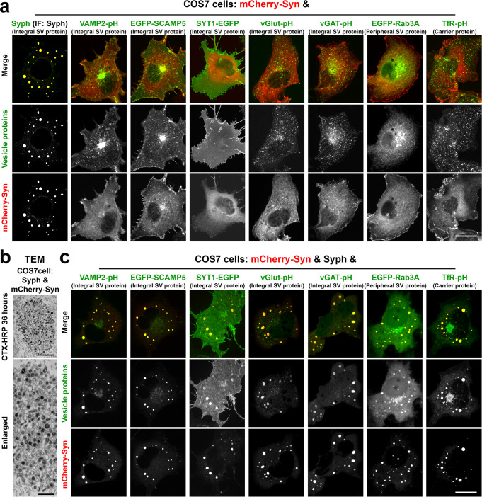
Fig. 1. Synaptophysin and synapsin condensates recruit other SV proteins.
a COS7 cells were co-transfected with mCherry-synapsin 1a (Syn) and one of these proteins as indicated: synaptophysin (Syph) (untagged), VAMP2-pHluorin (VAMP2-pH), EGFP-SCAMP5, synaptotagmin 1-EGFP (SYT1-EGFP), vesicular glutamate transporter 1-pHluorin (vGlut-pH), vesicular GABA transporter-pHluorin (vGAT-pH), EGFP-Rab3A, and transferrin receptor-pHluorin (TfR-pH). Synaptophysin, which was untagged, was revealed by immunofluorescence. Only cells that co-express mCherry-synapsin and synaptophysin show large droplets. b Synaptophysin and mCherry-synapsin expressing COS7 cells were exposed to 10 μg/ml cholera toxin conjugated HRP (CTX-HRP) for 36 h, then fixed, processed for HRP reactivity and embedded for transmission electron microscopy (TEM). Black vesicles are the vesicles labeled by the endocytic tracer CTX-HRP. c COS7 cells were triple transfected with synaptophysin, mCherry-synapsin and one other fluorescent fusion protein as in field a. Note that all SV proteins and the transferrin receptor co-assemble into the droplets formed by synaptophysin and mCherry-synapsin. See also Supplementary Fig. 1. Scale bars, a = 20 μm, b = 500 mm (top) and 200 nm (bottom), c = 20 μm.

