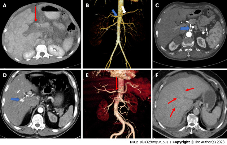
Figure 1.
Image examination. A: Axial computed tomography (CT) shows portal vein thrombosis; B: Separate origins of the common hepatic artery and the splenic artery. Coronal volume rendered image of a 44-year-old female presenting with right lumber pain demonstrates a hepatic arterial variation as the common hepatic artery (blue arrow) and the splenic artery (white arrow) originating directly from the abdominal aorta separately; C: The common hepatic artery directly originating from the abdominal aorta. Axial contrast-enhanced CT of a 55-year-old male patient with known peripheral vascular disease presenting direct origin of the common hepatic artery (arrow) from the abdominal aorta; D: Hepatic artery pseudoaneurysm. Axial contrast-enhanced arterial phase CT image of a 42-year-old man, suffering from epigastric pain and presenting with hemobilia and elevated liver enzymes, shows an 8 mm diameter pseudoaneurysm (arrow); E: High grade stenosis of the common hepatic artery. Coronal volume rendered CT image of a 76-year-old male with the diagnosis of vasculitis demonstrates severe stenosis (arrow); F: Occlusion of all hepatic veins consistent with Budd-Chiari syndrome. Venous phase axial CT scan shows thrombosed hepatic veins (arrows).

