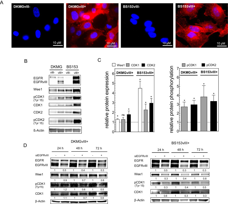
Fig. 1.
Wee1 expression in EGFRvIII− and EGFRvIII+ GBM cells. A EGFRvIII-specific immunofluorescence staining of DKMGvIII− /+ and BS153vIII− /+ cells. B Expression respectively phosphorylation of Wee1, CDK1 and CDK2 in DKMGvIII− /+ and BS153vIII− /+ cells. For Western blot analysis, samples were normalized to cell number. β-Actin served as loading control. C For quantification of protein expression and phosphorylation, the relative expression/phosphorylation values of EGFRvIII+ cells were normalized to the relative values of EGFRvIII− cells (n = 4; mean with S.E.M; p-values are obtained by Mann Whitney test, *p < 0.05). D Impact of siRNA-mediated EGFRvIII knockdown in DKMGvIII+ and BS153vIII+ cells on Wee1 and CDK expression and CDK1 phosphorylation after 24 h, 48 h and 72 h. An siRNA against cyclophilin B served as a control

