
Fig. 1.
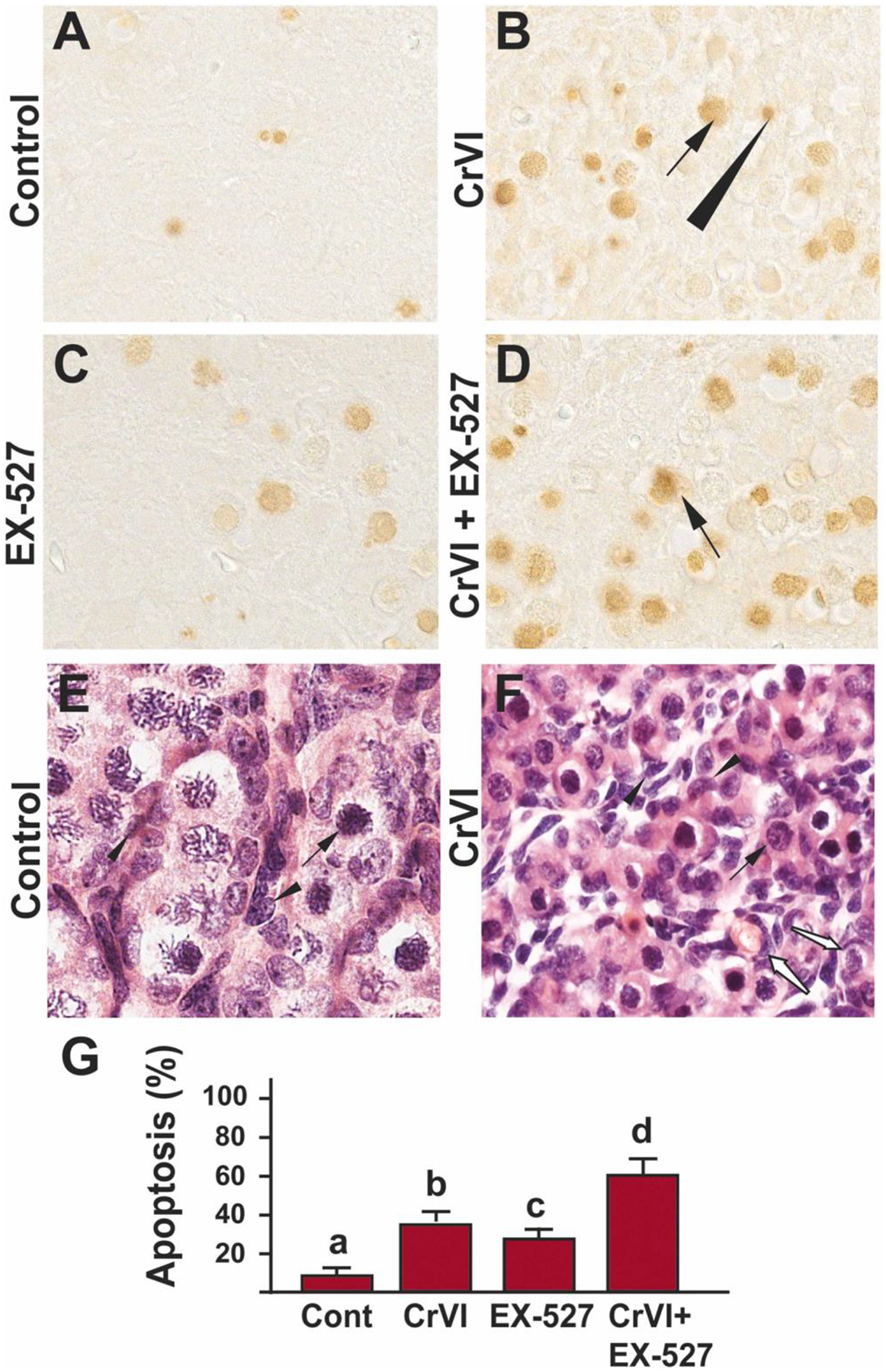
SIRT1 inhibitor exacerbated Cr(VI)-induced germ cell apoptosis of F1 offspring. Pregnant dams (F0) were exposed to Cr(VI) (10 ppm) through drinking water from 9.5–14.5 days post-coitum (dpc). Cr(VI) exposed and unexposed dams were injected (i.p.) with EX-527 (50 mg/kg body weight). On postnatal day (PND) 1, F1 offspring were euthanized, and ovaries were processed for TUNEL assay as described under Materials and methods. Representative images of the ovaries are shown from control (A), Cr(VI) (B), EX-527 (C), and Cr(VI)+EX-527 (D). H&E images of control (E) and Cr(VI)-exposed (F) ovaries are shown. The histogram shows the percentage of TUNEL-positive apoptotic cells (G). Different lowercase letters (a, b, c, d) indicate significant differences between groups (p<0.05). Each value is mean ± SEM 5 F0 rats (n=5). Arrows (germ cells) and arrowhead (somatic cell) indicate apoptotic TUNEL-positive cells, white arrows (F) show partly formed primordial follicles.