
Fig. 5.
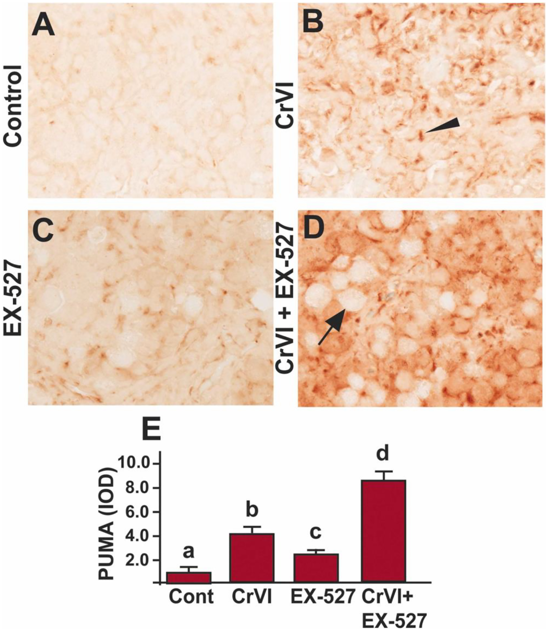
SIRT1 inhibitor increased PUMA in the ovary of F1 offspring. Pregnant dams (F0) were exposed to Cr(VI) (10 ppm) through drinking water from 9.5–14.5 days post-coitum (dpc). Cr(VI) exposed and unexposed dams were injected (i.p.) with EX-527 (50 mg/kg body weight). On postnatal day (PND) 1, F1 offspring were euthanized, and ovaries were processed for IHC. Representative images of the ovaries are shown from control (A), Cr(VI) (B), EX-527 (C), and Cr(VI)+EX-527 (D). The histogram shows the integrated optical density (IOD) (E). Different lowercase letters (a, b, c, d) indicate significant differences between groups (p<0.05). Each value is mean ± SEM 5 F0 rats (n=5). Arrow indicates germ cell, and the arrowhead indicates a somatic cell.