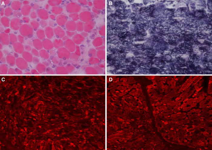
Figure 2.
Histochemical and immunohistochemical staining of muscle biopsies (×200). Following HE staining (A), different sizes of muscle fibers were observed, and degeneration, necrosis, and regeneration of the muscle fibers was evident as well as the deposition of amorphous materials. Some muscle fiber staining was significantly darker than the surrounding myofibrils according to modified Gömöri trichrome staining (B). Fluorescence antibody staining of desmin, α-crystallin (C), and myotilin (D) revealed high expression levels of these proteins in the sediment.

