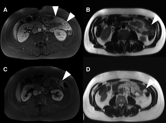
Figure 1.

Radiological presentation: (A) and (B) representative MRI images (A=T1 VIBE fat-saturated images postcontrast B=T2 haste) of the intraabdominal manifestations present in our patient before initiation of therapy with prednisone and MTX (C) and (D) follow-up MRI images after approx. 1 month of therapy with prednisone and MTX. (C=T1 VIBE fat-saturated images postcontrast D=T2 HASTE) The lesions (marked with Δ) show strong contrast uptake and are T2 hypointense. Additional diffusion weighted sequences (not shown) show no significant restriction of diffusion. MTX, methotrexate.
