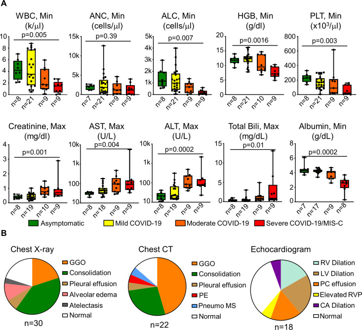
Figure 2.
Laboratory and imaging findings in CAR T-cell recipients with COVID-19. (A) Laboratory findings in asymptomatic SARS-CoV-2 infections, mild COVID-19, moderate COVID-19, and severe COVID-19/MIS-C groups. Min or max laboratory values over the course of illness are reported. Bars show the median and 25th–75th IQR. Comparisons between groups are with Kruskal-Wallis test with significance of p<0.05. (B) Distribution of abnormal imaging findings on chest X-ray, chest CT, and echocardiogram is demonstrated. ALT, alanine transaminase; ANC, absolute neutrophil count; AST, aspartate aminotransferase; bili, bilirubin; CA, coronary artery; GGO, ground-glass opacity; Hgb, hemoglobin; LV, left ventricular; max, maximum; min, minimum; MIS-C, multisystem inflammatory syndrome in children; PE, pulmonary embolism; PLT, platelet count; pneumo MS, pneumomediastinum; n, number of values; RV, right ventricular; RVP, right ventricular pressure; WBC, white blood cell.

