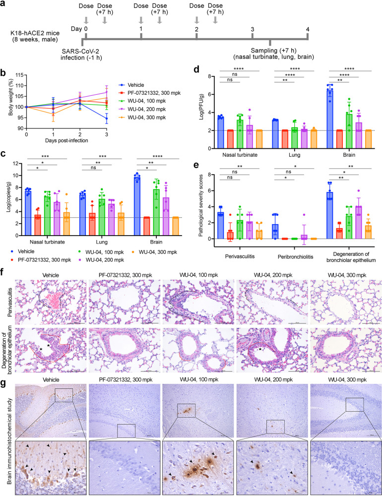
Figure 6.
In vivo efficacy of WU-04 against a SARS-CoV-2 in K18-hACE2 mice. (a) Schematic diagram of the study process. (b) Changes of the mouse body weights during the study. (c, d) After 3 days treatment with WU-04, PF-07321332, or the vehicle control, the viral RNA copy numbers (c) and the viral titers (d) in the nasal turbinate, lung, and brain of each mouse were determined. (e–g) After 3 days treatment, the lung tissues were harvested and processed for histological analysis (f) and the pathological changes were scored (e), and the brain tissues were harvested and processed for immunohistochemical analysis (g) using methods in the Supporting Information. Severe lymphoplasmacytic perivasculitis and vasculitis (black dotted line), peribronchiolar inflammatory infiltrate (red dotted line), degeneration of bronchiolar epithelium (asterisk), and viral antigen-positive cells in the brain (arrow) were marked. * P < 0.05, ** P < 0.01, *** P < 0.001, ***** P < 0.0001.

