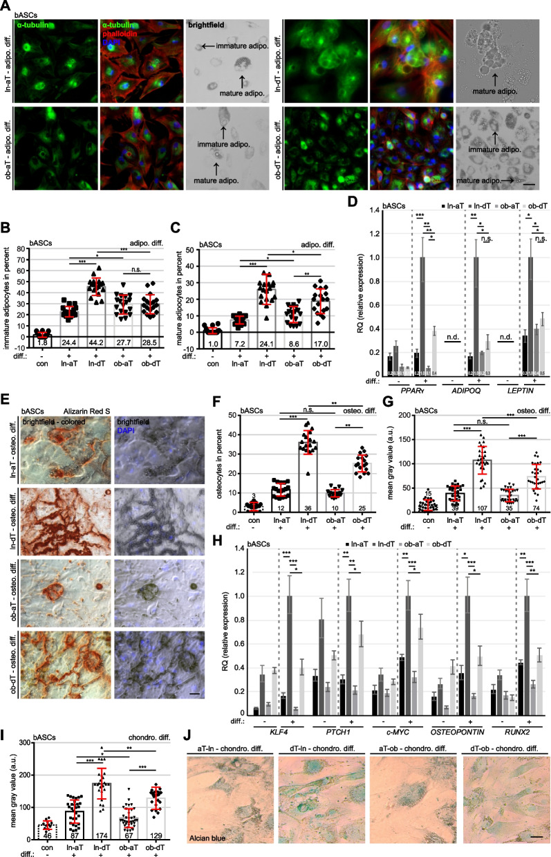
Fig. 1.
Lean and obese bASCs adjacent to breast cancer display an impaired differentiation capacity. A-J bASCs (ln-aT, ln-dT, ob-aT and ob-dT) were induced to adipogenic (adipo. Diff.) (A-D) for 14 days, osteogenic (osteo. Diff.) (E-H) and chondrogenic differentiation (chondro. Diff.) (I and J) for 21 days and their differentiation rates were evaluated. ln-dT bASCs were used as control cells without differentiation medium. A bASCs were stained for α-tubulin (green), phalloidin (red) to visualize the cytoskeleton, and DNA (DAPI, blue). Example images are shown. Scale bar: 50 μm. B and C The percentage of immature adipocytes (lipid vacuoles < 5 nm) (B) or mature adipocytes (lipid vacuoles > 5 nm) (C) was quantified. The results of individual bASC subgroups are presented as mean ± SEM (n = 500 cells for each condition, pooled from three experiments). D The gene expression of PPARγ, ADIPOQ and LEPTIN is shown for undifferentiated (−) and differentiated (+) bASCs. The results are from three individual experiments and presented as mean ± SEM. E bASCs were stained with Alizarin Red S to visualize calcium deposition. Representative images are shown. Scale bar: 50 μm. F and G The percentage of bASCs showing calcium deposition (F) and the mean gray value (G) were evaluated. The results are presented as mean ± SEM (F: n = 500 cells for each condition, pooled from three experiments, G: n = 30 images for each condition, pooled from three experiments). H The gene expression of KLF4, PTCH1, c-MYC, OSTEOPONTIN and RUNX2 is shown for undifferentiated (−) and differentiated (+) bASCs. The results are from three individual experiments and presented as mean ± SEM. I and J bASCs were stained with Alcian blue to visualize acidic polysaccharides. Representative bright-field images are shown (J). Scale bar: 50 μm. The quantification of the mean gray value is presented (I). The results are shown as mean ± SEM (n = 30 images for each condition, pooled from three experiments). Unpaired Mann-Whitney U test was used in (B and C), (F and G) and (I). ∗p < 0.05, ∗∗p < 0.01, ∗∗∗p < 0.001. Student’s t test was used in (D) and (H). ∗p < 0.05, ∗∗p < 0.01, ∗∗∗p < 0.001

