
Fig. 4. C. acnes triggers adipocyte differentiation and Camp expression in dermal fibroblasts.
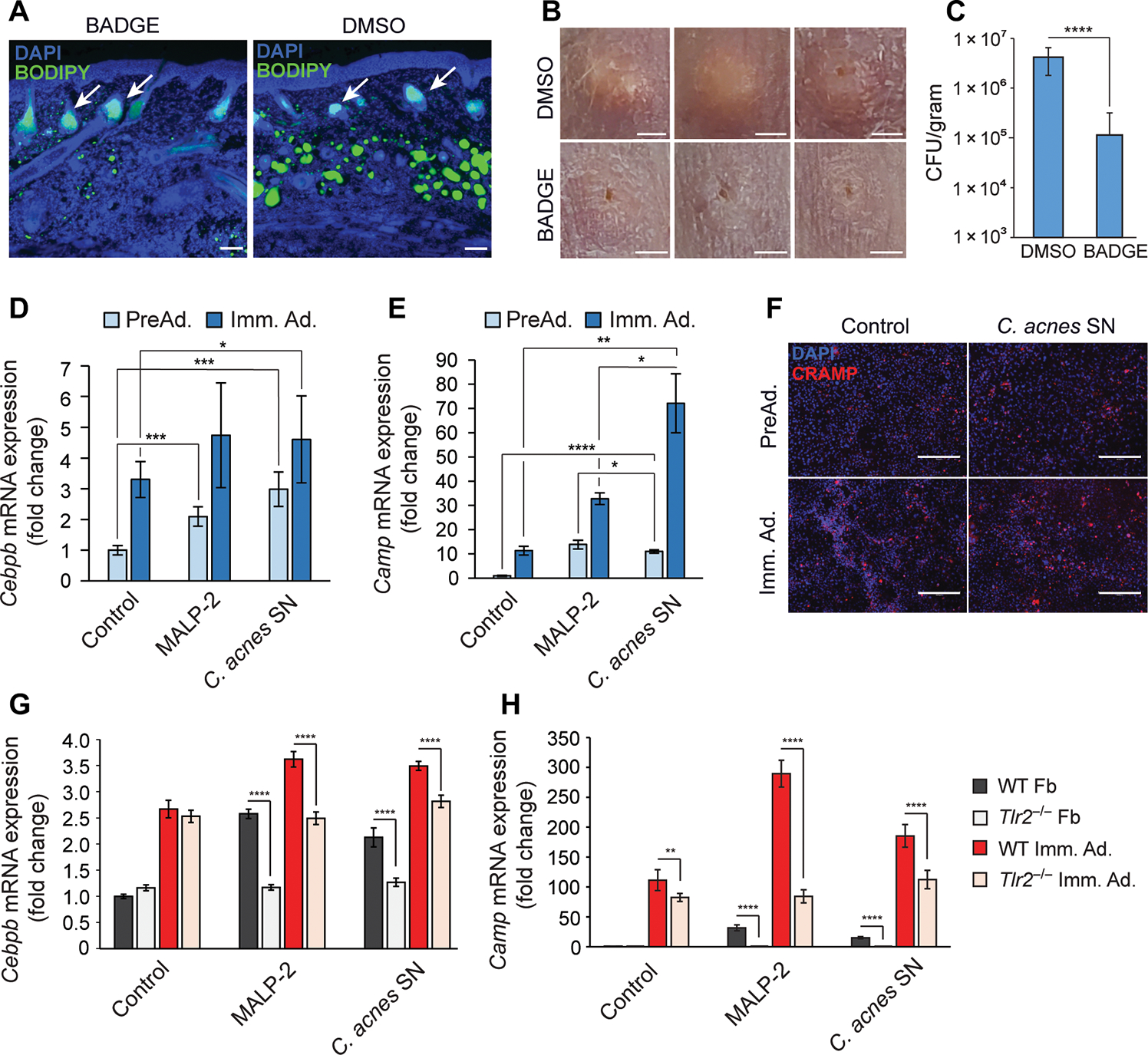
(A to C) SKH-1 mice were intraperitoneally injected daily with BADGE or vehicle (DMSO) as a control starting 1 day before intradermal infection with C. acnes. On day 6, skin was collected for analysis (n = 5). (A) Skin was stained with DAPI to stain nuclei or BODIPY to stain for the presence of lipid droplets. White arrows indicate sebaceous glands. Scale bar, 100 μm. (B) Representative images of the PBS control or C. acnes injection sites on mouse back after BADGE or DMSO vehicle treatment. Scale bar, 2 mm. (C) Enumeration of bacterial CFU from infected and mock-infected skin lesions of the infected area. (D and E) 3T3-L1 preadipocytes were cultured as nondifferentiated preadipocytes (PreAd.) or differentiated into immature adipocytes (Imm. Ad.) and stimulated for 24 hours with MALP-2 (100 ng/ml), 2.5% sterile conditioned C. acnes supernatant (SN), or bacterial culture medium (RCM) as control, and the relative mRNA expression of Cebpb (D) or Camp (E) was assessed by real-time qPCR. (F) Undifferentiated 3T3-L1 preadipocytes were stimulated with C. acnes SN for 48 hours, immunostained for CRAMP (red), and counterstained with DAPI (blue). Scale bar, 300 μm. (G and H) Primary dermal fibroblasts (Fb) isolated from wild-type (WT) or TLR2 knockout (Tlr2−/−) mice were stimulated with MALP-2 (100 ng/ml), 2.5% C. acnes SN, or RCM control for 24 hours in the presence or absence of differentiation medium, and the relative mRNA expression of Cebpb (G) or Camp (H) was assessed by real-time qPCR. All data are means ± SD. *P < 0.05, **P < 0.01, ***P < 0.001, and ****P < 0.0001 using Student’s paired t test.