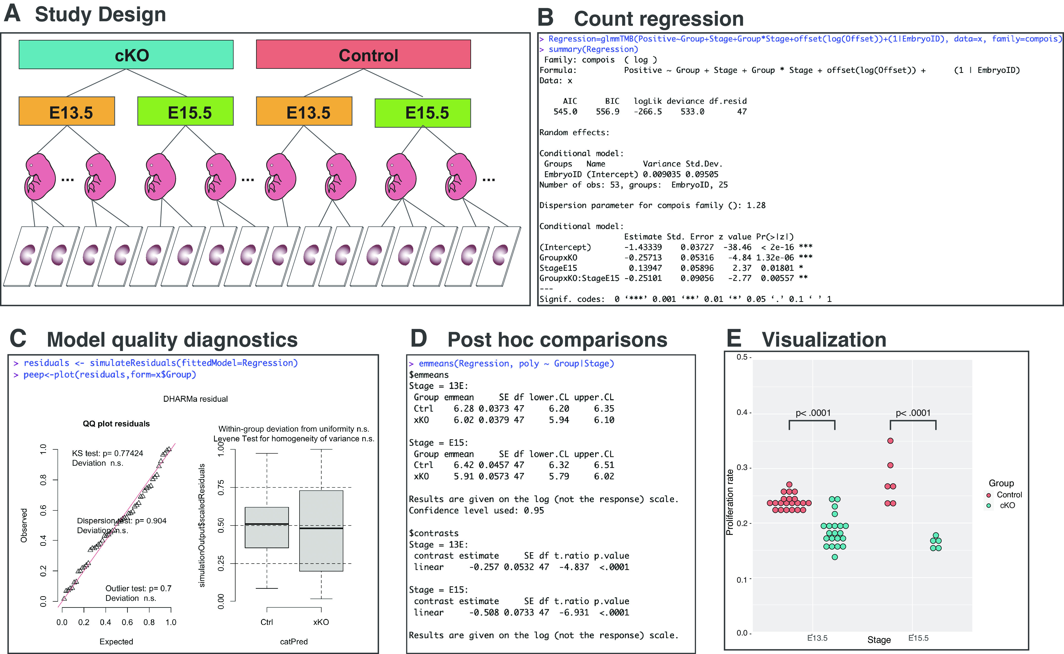
Figure 2.

Statistical analysis of cardiomyocyte proliferation data set. Counts of total and proliferating cardiomyocytes were retrieved from 53 microscopy slides from 25 embryonic hearts harvested at 2 embryonic stages (E13.5 and E15.5) belonging to 2 genotypes (control and cKO) (A). Rates of Edu+ cardiomyocytes were modeled using COM-Poisson regression as combination of experimental factors, random factor, and offset using glmmTMB package in R (B). The suitability of produced regression model was assessed with DHARMa by evaluating the variance and distribution of residuals (C). Post hoc comparison of proliferating cardiomyocyte rates between genotypes within each embryonic stage was performed using estimated marginal means package emmeans (D) and data visualized using ggplot2 (E). cKO, cardiomyocyte-specific knockout animal; COM, Conway-Maxwell.
