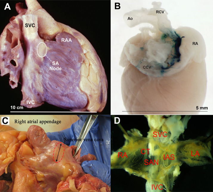
Figure 1.
Gross anatomy of human and mouse sinoatrial node (SAN). A: lateral view of a human heart specimen showing location of the SAN. Reproduced from Hayes et al. (5). B: posterior view of a X-Gal-stained mouse heart showing the SA nodal area. Reproduced from Lee et al. (6). C: dissected human heart showing SAN area (dotted circle). Reproduced from Mitrofanova et al. (7). D: dissected SAN tissue from a mouse. Approximate sizes are shown. Ao, aorta; CCV, common carotid vein; CT, crista terminalis; IAS, interatrial septum; IVC, inferior vena cava; LA, left atrium; RCV, right carotid vein; RA, right atrium; RAA, right atrial appendage; SVC, superior vena cava. Reproduced images, as indicated, are used with permission.

