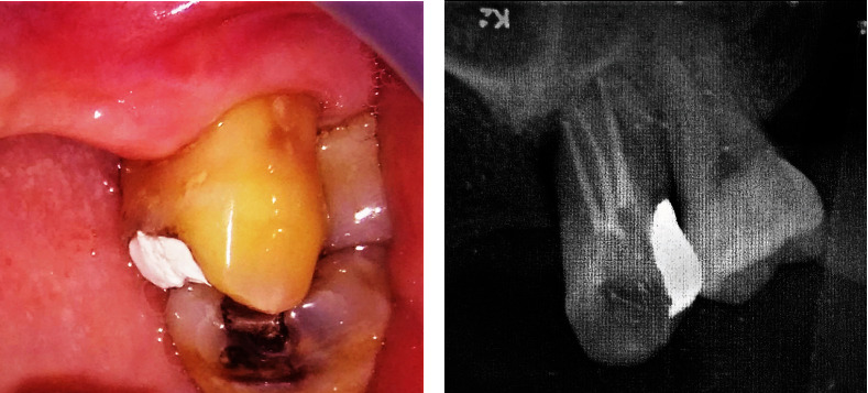
Figure 2.

Clinical and radiographic examination of tooth #26 after root canal treatment. (a) Temporary filling covered the mesio-occlusal part of crown tooth #26. Gingiva looked normal. (b) Radiographic examination showed obturation of root canals of tooth #26 with decreased radiolucency on the apical part of the tooth.
