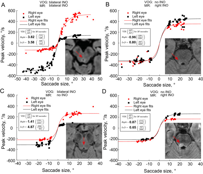
Fig. 3.
Concordance between VOG saccade measurements and MRI ratings found in four example MS patients. A Patient with bilateral INO. VOG measures and MR raters largely agreed, with two reviewers diagnosing a bilateral MLF lesion and one reviewer a unilateral left lesion. B VOG measurements detect no INO, but all MR reviewers found a right MLF lesion. C Patient with bilateral INO on VOG measures, but no MLF lesion detected by any MR rater. Rightward saccades indicate a clear left INO (VDI 4.87). The right INO is not very pronounced, but still significant (VDI 1.41). D All MR raters found a right MLF lesion, though VOG measurements do not confirm any INO. Note, however, that the peak velocities of all saccades appear abnormally low

