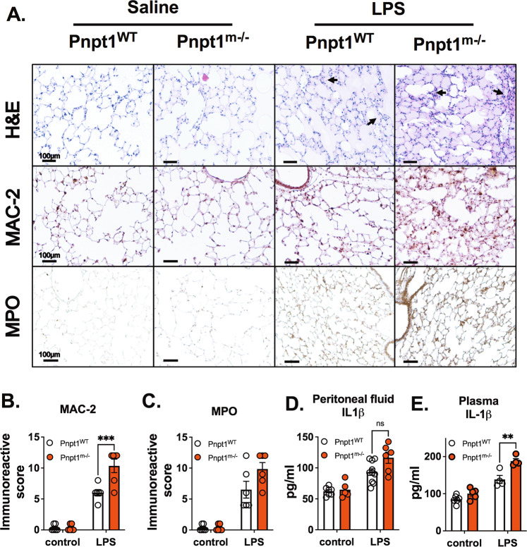
Fig. 1.
Pnpt1 deletion increased LPS-induced lung and systemic inflammation in vivo. Ten-week-old male Pnpt1m−/− and WT mice were sacrificed 6 h after i.p. injection of 40 mg/kg LPS or saline (control). Whole blood samples were collected, and the peritoneal cavities were washed with PBS. A Representative H&E, Mac-2, and MPO staining of lung sections. B Quantification of Mac-2 staining. C Quantification of MPO staining. ELISA analysis of IL-1β levels in (D) peritoneal lavage fluid and (E) plasma. Statistical analyses in (B–E) were performed using a 2-way ANOVA and Bonferroni’s post hoc test. N = 6 mice per group. ****P < 0.001 between the LPS-WT and LPS-Pnpt1m−/− groups. Bars represent the mean ± SEM. **p < 0.01, ***p < 0.001

