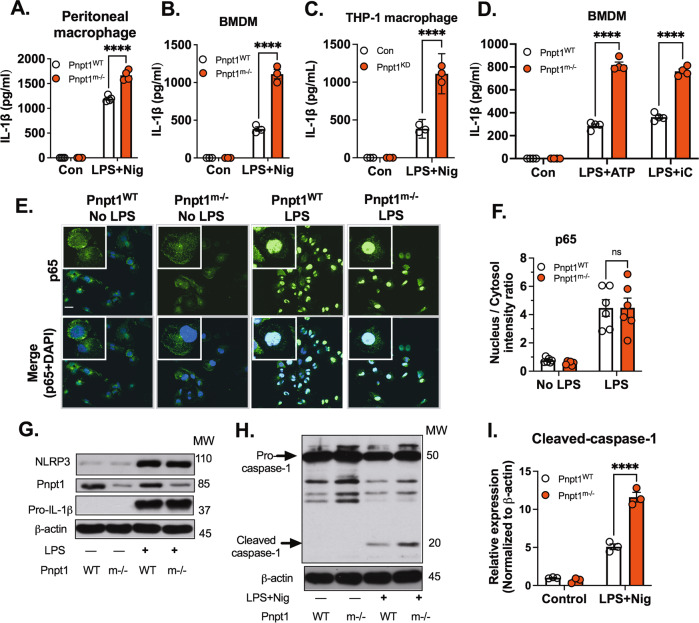
Fig. 3.
Pnpt1 deletion increased inflammasome activation independent of the NF-ĸB pathway. Macrophages from WT and Pnpt1m−/− mice were stimulated with or without LPS (100 ng/mL) for 3 h, followed by 2 µM nigericin (Nig) stimulation for 1 h. IL-1β release in the medium of (A) peritoneal macrophages, (B) BMDMs, and (C) THP-1 macrophages (control and Pnpt1 knockdown). BMDMs from WT and Pnpt1m−/− mice were stimulated with or without LPS (100 ng/mL) for 3 h and with (D) 2 µM ATP for 1 h. 10 µg/mL poly (I:C) for 24 h. IL-1β release in the medium was measured by ELISA. (Con: control). E Peritoneal macrophages were stimulated with LPS (100 ng/mL) for 1 h. NF-κB p65 translocation was measured by immunofluorescence (scale bar: 10 µm), DAPI (blue), and p65 (green). F Quantification was performed by ImageJ. N = 3 experiments. G Peritoneal macrophages were stimulated with or without LPS (100 ng/mL) for 3 h. The protein expression of NLRP3 and pro-IL-1β was analyzed by western blotting, and the images are representative of three independent experiments. H, I Peritoneal macrophages were stimulated with LPS (100 ng/mL) for 3 h and with 1 µM nigericin (Nig) for 30 min. The western blots are representative of three independent experiments. Statistical analyses in (A–D, F and I) were performed using a 2-way ANOVA and Bonferroni’s post hoc test. ****P < 0.001 between the Pnpt1m−/− and WT groups after stimulation. Bars represent the mean ± SEM

