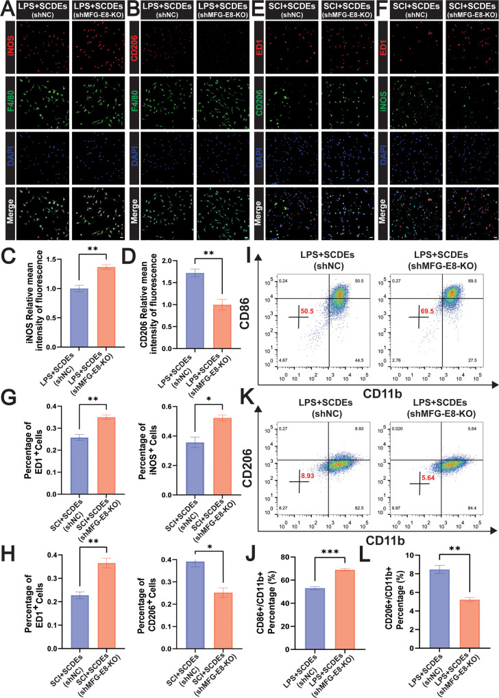
Fig. 7. MFG-E8 knockout suppressed the M2 polarization in vitro and vivo.
A Representative immunofluorescence staining images of iNOS (red) and F4/80 (green) in vitro. Nuclei were labeled with DAPI (blue) in each group (n = 5). Scale bar: 20 μm. B Representative immunofluorescence staining images of CD206 (red) and F4/80 (green) in vitro. Nuclei were labeled with DAPI (blue) in each group (n = 5). Scale bar: 20 μm. C, D The results of the iNOS and CD206 relative mean intensity of the fluorescence (n = 5). E Representative immunofluorescence staining images of iNOS (green) and ED1 (red) in vivo. Nuclei were labeled with DAPI (blue) in each group (n = 3). Scale bar: 20 μm. F Representative immunofluorescence staining images of CD206 (green) and ED1 (red) in vivo. Nuclei were labeled with DAPI (blue) in each group (n = 3). Scale bar: 20 μm. G, H Quantitative analysis of the positive cells of ED1, iNOS, and CD206 (n = 3). I, J Flow cytometry assay detected CD86+/CD11b + M1 phenotype, indicating that MFG-E8 knockout promoted the M1 polarization (n = 3). K, L Flow cytometry assay detected CD206+/CD11b + M2 phenotype, showing that MFG-E8 knockout inhibited the M2 polarization (n = 3). Data were presented as mean ± SEM. Results were analyzed by One-way ANOVA. Significance: *P < 0.05, **P < 0.01, ***P < 0.001.

