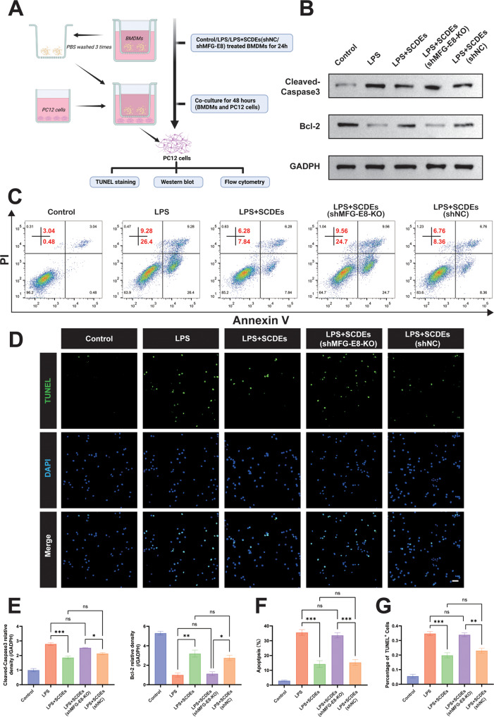
Fig. 8. Improved inflammatory microenvironment inhibited neuronal apoptosis.
A Schematic representation of co-culture of PC12 cells and BMDMs. B, E Representative western blots of Cleaved Caspase-3 and Bcl-2 in PC12 cells. Quantitative analysis of the Cleaved Caspase-3/GAPDH ratio and Bcl-2/GAPDH ratio in PC12 cells (n = 3). C, F Flow cytometry assay detected apoptosis of PC12 cells in each group, showing that SCDEs inhibited LPS-induced apoptosis and that the knockout of MFG-E8 reversed the positive effect (n = 3). D, G The TUNEL staining of PC12 cells showed that SCDEs reduced TUNEL + cells while MFG-E8 knockout increased TUNEL + cells (n = 3). Scale bar: 50 μm. Data were presented as mean ± SEM. Results were analyzed by One-way ANOVA. Significance: ns-not significant, *P < 0.05, **P < 0.01, ***P < 0.001.

