Figure 1.
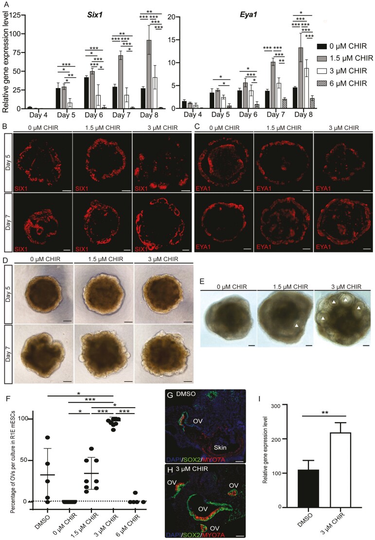
Early Wnt modulation improves inner ear organoid induction. (A) Mean (±SD) expression levels of 2 pre-placodal ectoderm (PPE) marker genes, Six1 and Eya1, under various Wnt levels during the early induction of inner ear organoids (n = 4-6 independent differentiation experiments). Two-way ANOVA: day*** and treatment***. Representative immunohistochemistry (IHC) images of SIX1 (B) and EYA1 (C) signals at the outer layer of samples under various Wnt levels on D5 and 7. (D) Representative bright field (BF) images of aggregates on D5 and D7. (E) Representative BF images of aggregates on D10. More prominent otic vesicles (OVs; white triangles) were seen in 3 µM CHIR samples. (F) Significant increase in the production of OVs in 3 µM CHIR samples. Each dot indicates one 96 well-plate experiment (n = 5-8 independent differentiation experiments). One-way ANOVA of treatment (P = .005) followed by post hoc tests. (G-H) Representative IHC images of D20 OVs in the DMSO control and 3 µM CHIR treatment. (I) Mean (±SD) gene expression levels of the hair cell marker myosin 7A in D20 DMSO controls and the 3 µM CHIR treatment (n = 3 independent differentiation experiments). Scale bars: 100 µm. *, **, and *** denote P < .05, <.001, and <.0001, respectively.

